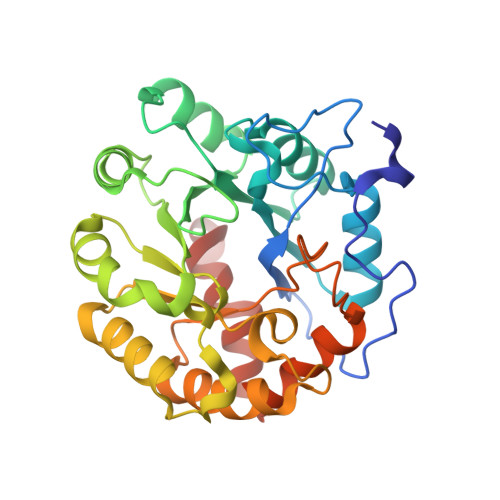
4DI9

CRYSTAL STRUCTURE OF THE D248A mutant of 2-PYRONE-4,6-DICARBOXYLIC ACID HYDROLASE FROM SPHINGOMONAS PAUCIMOBILIS complexed with substrate at pH 6.5


Experimental Data Snapshot

  • Method: X-RAY DIFFRACTION
  • Resolution: 1.35 Å
  • R-Value Free: 0.183 
  • R-Value Work: 0.160 
  • R-Value Observed: 0.161 

wwPDB Validation   3D Report Full Report


Ligand Structure Quality Assessment 


This is version 1.5 of the entry. See complete history


Literature

Structure and Catalytic Mechanism of LigI: Insight into the Amidohydrolase Enzymes of cog3618 and Lignin Degradation.

Hobbs, M.E.Malashkevich, V.Williams, H.J.Xu, C.Sauder, J.M.Burley, S.K.Almo, S.C.Raushel, F.M.

(2012) Biochemistry 51: 3497-3507

  • DOI: https://doi.org/10.1021/bi300307b
  • Primary Citation of Related Structures:  
    4D8L, 4DI8, 4DI9, 4DIA

  • PubMed Abstract: 

    LigI from Sphingomonas paucimobilis catalyzes the reversible hydrolysis of 2-pyrone-4,6-dicarboxylate (PDC) to 4-oxalomesaconate and 4-carboxy-2-hydroxymuconate in the degradation of lignin. This protein is a member of the amidohydrolase superfamily of enzymes. The protein was expressed in Escherichia coli and then purified to homogeneity. The purified recombinant enzyme does not contain bound metal ions, and the addition of metal chelators or divalent metal ions to the assay mixtures does not affect the rate of product formation. This is the first enzyme from the amidohydrolase superfamily that does not require a divalent metal ion for catalytic activity. The kinetic constants for the hydrolysis of PDC are 340 s(-1) and 9.8 × 10(6) M(-1) s(-1) (k(cat) and k(cat)/K(m), respectively). The pH dependence on the kinetic constants suggests that a single active site residue must be deprotonated for the hydrolysis of PDC. The site of nucleophilic attack was determined by conducting the hydrolysis of PDC in (18)O-labeled water and subsequent (13)C nuclear magnetic resonance analysis. The crystal structures of wild-type LigI and the D248A mutant in the presence of the reaction product were determined to a resolution of 1.9 Å. The C-8 and C-11 carboxylate groups of PDC are coordinated within the active site via ion pair interactions with Arg-130 and Arg-124, respectively. The hydrolytic water molecule is activated by the transfer of a proton to Asp-248. The carbonyl group of the lactone substrate is activated by electrostatic interactions with His-180, His-31, and His-33.


  • Organizational Affiliation

    Department of Biochemistry and Biophysics, Texas A&M University, College Station, TX 77843, USA.


Macromolecules
Find similar proteins by:  (by identity cutoff)  |  3D Structure
Entity ID: 1
MoleculeChains Sequence LengthOrganismDetailsImage
2-pyrone-4,6-dicarbaxylate hydrolase303Sphingomonas paucimobilisMutation(s): 1 
Gene Names: ligI
UniProt
Find proteins for O87170 (Sphingobium sp. (strain NBRC 103272 / SYK-6))
Explore O87170 
Go to UniProtKB:  O87170
Entity Groups  
Sequence Clusters30% Identity50% Identity70% Identity90% Identity95% Identity100% Identity
UniProt GroupO87170
Sequence Annotations
Expand
  • Reference Sequence
Small Molecules
Ligands 2 Unique
IDChains Name / Formula / InChI Key2D Diagram3D Interactions
0GY
Query on 0GY

Download Ideal Coordinates CCD File 
B [auth A](1E,3Z)-4-hydroxybuta-1,3-diene-1,2,4-tricarboxylic acid
C7 H6 O7
QWLUKZXOQAQUFQ-NAOWAUKJSA-N
ACT
Query on ACT

Download Ideal Coordinates CCD File 
C [auth A]ACETATE ION
C2 H3 O2
QTBSBXVTEAMEQO-UHFFFAOYSA-M
Experimental Data & Validation

Experimental Data

  • Method: X-RAY DIFFRACTION
  • Resolution: 1.35 Å
  • R-Value Free: 0.183 
  • R-Value Work: 0.160 
  • R-Value Observed: 0.161 
  • Space Group: C 1 2 1
Unit Cell:
Length ( Å )Angle ( ˚ )
a = 73.552α = 90
b = 50.952β = 91.86
c = 73.346γ = 90
Software Package:
Software NamePurpose
SCALEPACKdata scaling
PHASERphasing
REFMACrefinement
PDB_EXTRACTdata extraction
CBASSdata collection
HKL-3000data reduction

Structure Validation

View Full Validation Report


Ligand Structure Quality Assessment 


Entry History 

Deposition Data

Revision History  (Full details and data files)

  • Version 1.0: 2012-10-03
    Type: Initial release
  • Version 1.1: 2013-01-09
    Changes: Database references
  • Version 1.2: 2013-01-23
    Changes: Database references
  • Version 1.3: 2013-02-20
    Changes: Database references
  • Version 1.4: 2021-02-10
    Changes: Database references, Derived calculations
  • Version 1.5: 2024-02-28
    Changes: Data collection, Database references