4DFY | pdb_00004dfy

Crystal structure of R194A mutant of cAMP-dependent protein kinase with unphosphorylated activation loop


Experimental Data Snapshot

  • Method: X-RAY DIFFRACTION
  • Resolution: 3.00 Å
  • R-Value Free: 
    0.286 (Depositor), 0.284 (DCC) 
  • R-Value Work: 
    0.252 (Depositor), 0.245 (DCC) 
  • R-Value Observed: 
    0.253 (Depositor) 

Starting Model: experimental
View more details

wwPDB Validation 3D Report Full Report

Validation slider image for 4DFY

This is version 1.4 of the entry. See complete history

Literature

Structural basis for the regulation of protein kinase a by activation loop phosphorylation.

Steichen, J.M.Kuchinskas, M.Keshwani, M.M.Yang, J.Adams, J.A.Taylor, S.S.

(2012) J Biological Chem 287: 14672-14680

  • DOI: https://doi.org/10.1074/jbc.M111.335091
  • Primary Citation Related Structures: 
    4DFY

  • PubMed Abstract: 

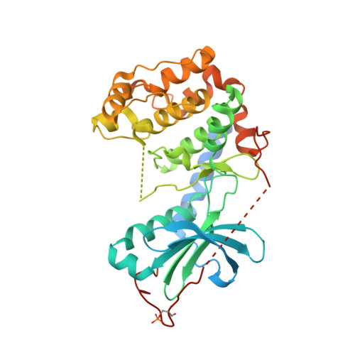
    The catalytic subunit of cAMP-dependent protein kinase (PKA) is a member of the AGC group of protein kinases. Whereas PKA has served as a structural model for the protein kinase superfamily, all previous structures of the catalytic subunit contain a phosphorylated activation loop. To understand the structural effects of activation loop phosphorylation at Thr-197 we used a PKA mutant that does not autophosphorylate at Thr-197. The enzyme crystallized in the apo-state, and the structure was solved to 3.0 Å. The N-lobe is rotated by 18° relative to the wild-type apoenzyme, which illustrates that the enzyme likely exists in a wide range of conformations in solution due to the uncoupling of the N- and C-lobes. Several regions of the protein including the activation loop are disordered in the structure, and there are alternate main chain conformations for the magnesium positioning loop and catalytic loop causing a complete loss of hydrogen bonding between these two active site structural elements. These alterations are reflected in a 20-fold decrease in the apparent phosphoryl transfer rate as measured by pre-steady-state kinetic methods.


  • Organizational Affiliation
    • Department of Chemistry and Biochemistry, University of California at San Diego, La Jolla, California 92093, USA.

Macromolecule Content 

  • Total Structure Weight: 85.59 kDa 
  • Atom Count: 4,991 
  • Modeled Residue Count: 624 
  • Deposited Residue Count: 742 
  • Unique protein chains: 1

Macromolecules

Find similar proteins by:|  3D Structure
Entity ID: 1
MoleculeChains  Sequence LengthOrganismDetailsImage
cAMP-dependent protein kinase catalytic subunit alphaA,
B [auth E]
371Mus musculusMutation(s): 1 
Gene Names: PrkacaPkaca
EC: 2.7.11.11
UniProt
Find proteins for P05132 (Mus musculus)
Explore P05132 
Go to UniProtKB:  P05132
Entity Groups
Sequence Clusters30% Identity50% Identity70% Identity90% Identity95% Identity100% Identity
UniProt GroupP05132
Sequence Annotations
Expand
Reference Sequence

Small Molecules

Modified Residues  1 Unique
IDChains TypeFormula2D DiagramParent
SEP
Query on SEP
A,
B [auth E]
L-PEPTIDE LINKINGC3 H8 N O6 PSER

Experimental Data & Validation

Experimental Data

  • Method: X-RAY DIFFRACTION
  • Resolution: 3.00 Å
  • R-Value Free:  0.286 (Depositor), 0.284 (DCC) 
  • R-Value Work:  0.252 (Depositor), 0.245 (DCC) 
  • R-Value Observed: 0.253 (Depositor) 
Space Group: P 32 2 1
Unit Cell:
Length ( Å )Angle ( ˚ )
a = 95.745α = 90
b = 95.745β = 90
c = 173.959γ = 120
Software Package:
Software NamePurpose
ADSCdata collection
PHASERphasing
REFMACrefinement
HKL-2000data reduction
SCALEPACKdata scaling

Structure Validation

View Full Validation Report


Entry History 

Deposition Data

Revision History  (Full details and data files)

  • Version 1.0: 2012-02-15
    Type: Initial release
  • Version 1.1: 2012-02-29
    Changes: Database references
  • Version 1.2: 2012-05-16
    Changes: Database references
  • Version 1.3: 2023-09-13
    Changes: Data collection, Database references, Derived calculations, Refinement description
  • Version 1.4: 2024-10-30
    Changes: Structure summary