Crystal Structure of Cell Adhesion Molecule Nectin-2/CD112 and Its Binding to Immune Receptor DNAM-1/CD226
Liu, J., Qian, X., Chen, Z., Xu, X., Gao, F., Zhang, S., Zhang, R., Qi, J., Gao, G.F., Yan, J.(2012) J Immunol 188: 5511-5520
- PubMed: 22547693 Search on PubMed
- DOI: https://doi.org/10.4049/jimmunol.1200324
- Primary Citation Related Structures:
4DFH, 4DFI - PubMed Abstract:
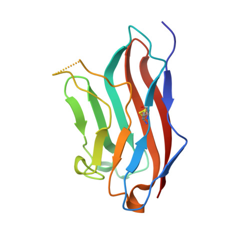
The nectin and nectin-like molecule (Necl) family includes important cell adhesion molecules (CAMs) characterized by their Ig-like nature. Such CAMs regulate a broad spectrum of cell-cell interactions, including the interaction between NK cells and cytotoxic T lymphocytes (CTLs) and their target cells. CAM members nectin-2 (CD112) and Necl-5 (CD155) are believed to form homodimers (for nectin-2) or heterodimers in their functions for cell adhesion, as well as to interact with immune costimulatory receptor DNAX accessory molecule 1 (DNAM-1) (CD226) to regulate functions of both NK and CTL cells. However, the structural basis of the interactive mode of DNAM-1 with nectin-2 or Necl-5 is not yet understood. In this study, a soluble nectin-2 Ig-like V-set domain (nectin-2v) was successfully prepared and demonstrated to bind to both soluble ectodomain and cell surface-expressed full-length DNAM-1. The 1.85-Å crystal structure of nectin-2v displays a perpendicular homodimer arrangement, revealing the homodimer characteristics of the nectin and Necls. Further mutational analysis indicated that disruption of the homodimeric interface of nectin-2v led to a failure of the homodimer formation, as confirmed by crystal structure and biochemical properties of the mutant protein of nectin-2v. Interestingly, the monomer mutant also loses DNAM-1 binding, as evidenced by cell staining with tetramers and surface plasmon resonance assays. The data indicate that interaction with DNAM-1 requires either the homodimerization or engagement of the homodimeric interface of nectin-2v. These results have implications for immune intervention of tumors or autoimmune diseases in the DNAM-1/nectin-2-dependent pathway.
- CAS Key Laboratory of Pathogenic Microbiology and Immunology, Institute of Microbiology, Chinese Academy of Sciences, Beijing, 100101 China.
Organizational Affiliation: