4D6W

Crystal Structure of the low pH conformation of Chandipura Virus glycoprotein G ectodomain


Experimental Data Snapshot

  • Method: X-RAY DIFFRACTION
  • Resolution: 3.60 Å
  • R-Value Free: 0.260 
  • R-Value Work: 0.193 
  • R-Value Observed: 0.197 

wwPDB Validation   3D Report Full Report


Ligand Structure Quality Assessment 


This is version 2.1 of the entry. See complete history


Literature

Structure of the Low Ph Conformation of Chandipura Virus G Reveals Important Features in the Evolution of the Vesiculovirus Glycoprotein.

Baquero, E.Albertini, A.A.Raux, H.Buonocore, L.Rose, J.K.Bressanelli, S.Gaudin, Y.

(2015) PLoS Pathog 11: 04756

  • DOI: https://doi.org/10.1371/journal.ppat.1004756
  • Primary Citation of Related Structures:  
    4D6W

  • PubMed Abstract: 

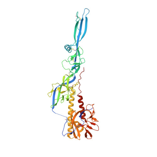
    Chandipura virus (CHAV), a member of the vesiculovirus genus, is an emerging human pathogen. As for other rhabdoviruses, CHAV entry into susceptible cells is mediated by its single envelope glycoprotein G which is both involved in receptor recognition and fusion of viral and cellular membranes. Here, we have characterized the fusion properties of CHAV-G. As for vesicular stomatitis virus (VSV, the prototype of the genus) G, fusion is triggered at low pH below 6.5. We have also analyzed the biochemical properties of a soluble form of CHAV-G ectodomain (CHAV-Gth, generated by thermolysin limited-proteolysis of recombinant VSV particles in which the G gene was replaced by that of CHAV). The overall behavior of CHAV-Gth is similar to that previously reported for VSV-Gth. Particularly, CHAV-Gth pre-fusion trimer is not stable in solution and low-pH-induced membrane association of CHAV-Gth is reversible. Furthermore, CHAV-Gth was crystallized in its low pH post-fusion conformation and its structure was determined at 3.6Å resolution. An overall comparison of this structure with the previously reported VSV-Gth post-fusion conformation, shows a high structural similarity as expected from the comparison of primary structure. Among the three domains of G, the pleckstrin homology domain (PHD) appears to be the most divergent and the largest differences are confined to the secondary structure of the major antigenic site of rhabdoviruses. Finally, local differences indicate that CHAV has evolved alternate structural solutions in hinge regions between PH and fusion domains but also distinct pH sensitive switches. Globally the comparison between the post fusion conformation of CHAV and VSV-G highlights several features essential for the protein's function. It also reveals the remarkable plasticity of G in terms of local structures.


  • Organizational Affiliation

    Institute for Integrative Biology of the Cell (I2BC), CEA, CNRS, Université Paris-Sud, Gif-sur-Yvette, France.


Macromolecules
Find similar proteins by:  (by identity cutoff)  |  3D Structure
Entity ID: 1
MoleculeChains Sequence LengthOrganismDetailsImage
GLYCOPROTEIN G
A, B, C
419Chandipura virusMutation(s): 0 
UniProt
Find proteins for P13180 (Chandipura virus (strain I653514))
Explore P13180 
Go to UniProtKB:  P13180
Entity Groups  
Sequence Clusters30% Identity50% Identity70% Identity90% Identity95% Identity100% Identity
UniProt GroupP13180
Sequence Annotations
Expand
  • Reference Sequence
Oligosaccharides

Help

Entity ID: 2
MoleculeChains Length2D Diagram Glycosylation3D Interactions
2-acetamido-2-deoxy-beta-D-glucopyranose-(1-4)-2-acetamido-2-deoxy-beta-D-glucopyranose
D
2N-Glycosylation
Glycosylation Resources
GlyTouCan:  G42666HT
GlyCosmos:  G42666HT
GlyGen:  G42666HT
Entity ID: 3
MoleculeChains Length2D Diagram Glycosylation3D Interactions
alpha-L-fucopyranose-(1-6)-2-acetamido-2-deoxy-beta-D-glucopyranose
E
2N-Glycosylation
Glycosylation Resources
GlyTouCan:  G86851RC
GlyCosmos:  G86851RC
GlyGen:  G86851RC
Small Molecules
Ligands 1 Unique
IDChains Name / Formula / InChI Key2D Diagram3D Interactions
NAG
Query on NAG

Download Ideal Coordinates CCD File 
F [auth B],
G [auth C]
2-acetamido-2-deoxy-beta-D-glucopyranose
C8 H15 N O6
OVRNDRQMDRJTHS-FMDGEEDCSA-N
Experimental Data & Validation

Experimental Data

  • Method: X-RAY DIFFRACTION
  • Resolution: 3.60 Å
  • R-Value Free: 0.260 
  • R-Value Work: 0.193 
  • R-Value Observed: 0.197 
  • Space Group: C 1 2 1
Unit Cell:
Length ( Å )Angle ( ˚ )
a = 365.59α = 90
b = 83.5β = 96.95
c = 60.82γ = 90
Software Package:
Software NamePurpose
PHENIXrefinement
XDSdata reduction
XSCALEdata scaling
PHASERphasing

Structure Validation

View Full Validation Report


Ligand Structure Quality Assessment 


Entry History 

Deposition Data

Revision History  (Full details and data files)

  • Version 1.0: 2015-03-04
    Type: Initial release
  • Version 1.1: 2015-04-08
    Changes: Database references
  • Version 1.2: 2019-03-06
    Changes: Data collection, Derived calculations, Experimental preparation, Other
  • Version 1.3: 2019-05-08
    Changes: Data collection, Experimental preparation
  • Version 2.0: 2020-07-29
    Type: Remediation
    Reason: Carbohydrate remediation
    Changes: Advisory, Atomic model, Data collection, Derived calculations, Other, Structure summary
  • Version 2.1: 2023-12-20
    Changes: Data collection, Database references, Derived calculations, Refinement description, Structure summary