Crystal Structure of Gib2, a Signal-Transducing Protein Scaffold Associated with Ribosomes in Cryptococcus Neoformans.
Ero, R., Dimitrova, V.T., Chen, Y., Bu, W., Feng, S., Liu, T., Wang, P., Xue, C., Tan, S.M., Gao, Y.G.(2015) Sci Rep 5: 8688
- PubMed: 25732347 Search on PubMedSearch on PubMed Central
- DOI: https://doi.org/10.1038/srep08688
- Primary Citation Related Structures:
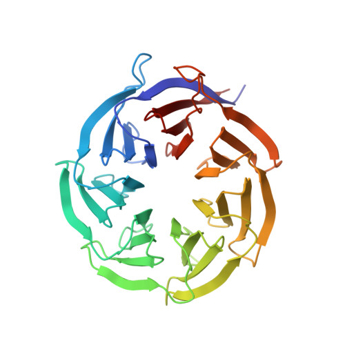
4D6V - PubMed Abstract:
The atypical Gβ-like/RACK1 Gib2 protein promotes cAMP signalling that plays a central role in regulating the virulence of Cryptococcus neoformans. Gib2 contains a seven-bladed β transducin structure and is emerging as a scaffold protein interconnecting signalling pathways through interactions with various protein partners. Here, we present the crystal structure of Gib2 at a 2.2-Å resolution. The structure allows us to analyse the association between Gib2 and the ribosome, as well as to identify the Gib2 amino acid residues involved in ribosome binding. Our studies not only suggest that Gib2 has a role in protein translation but also present Gib2 as a physical link at the crossroads of various regulatory pathways important for the growth and virulence of C. neoformans.
- School of Biological Sciences, Nanyang Technological University, Singapore.
Organizational Affiliation: