4D3L

SeMet structure of a novel carbohydrate binding module from glycoside hydrolase family 9 (Cel9A) from Ruminococcus flavefaciens FD-1 in the orthorhombic form


Experimental Data Snapshot

  • Method: X-RAY DIFFRACTION
  • Resolution: 2.00 Å
  • R-Value Free: 0.245 
  • R-Value Work: 0.212 
  • R-Value Observed: 0.214 

wwPDB Validation   3D Report Full Report


Ligand Structure Quality Assessment 


This is version 1.2 of the entry. See complete history


Literature

Complexity of the Ruminococcus Flavefaciens Cellulosome Reflects an Expansion in Glycan Recognition.

Venditto, I.Luis, A.S.Rydahl, M.Schuckel, J.Fernandes, V.O.Vidal-Melgosa, S.Bule, P.Goyal, A.Pires, V.M.R.Dourado, C.G.Ferreira, L.M.A.Coutinho, P.M.Henrissat, B.Knox, J.P.Basle, A.Najmudin, S.Gilbert, H.J.Willats, W.G.T.Fontes, C.M.G.A.

(2016) Proc Natl Acad Sci U S A 113: 7136

  • DOI: https://doi.org/10.1073/pnas.1601558113
  • Primary Citation of Related Structures:  
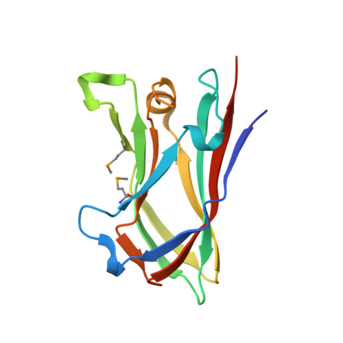
    4D3L, 4V17, 4V18, 4V1B, 4V1I, 4V1K, 4V1L, 5AOS, 5AOT, 5FU2, 5FU3, 5FU4, 5FU5

  • PubMed Abstract: 

    The breakdown of plant cell wall (PCW) glycans is an important biological and industrial process. Noncatalytic carbohydrate binding modules (CBMs) fulfill a critical targeting function in PCW depolymerization. Defining the portfolio of CBMs, the CBMome, of a PCW degrading system is central to understanding the mechanisms by which microbes depolymerize their target substrates. Ruminococcus flavefaciens, a major PCW degrading bacterium, assembles its catalytic apparatus into a large multienzyme complex, the cellulosome. Significantly, bioinformatic analyses of the R. flavefaciens cellulosome failed to identify a CBM predicted to bind to crystalline cellulose, a key feature of the CBMome of other PCW degrading systems. Here, high throughput screening of 177 protein modules of unknown function was used to determine the complete CBMome of R. flavefaciens The data identified six previously unidentified CBM families that targeted β-glucans, β-mannans, and the pectic polysaccharide homogalacturonan. The crystal structures of four CBMs, in conjunction with site-directed mutagenesis, provide insight into the mechanism of ligand recognition. In the CBMs that recognize β-glucans and β-mannans, differences in the conformation of conserved aromatic residues had a significant impact on the topology of the ligand binding cleft and thus ligand specificity. A cluster of basic residues in CBM77 confers calcium-independent recognition of homogalacturonan, indicating that the carboxylates of galacturonic acid are key specificity determinants. This report shows that the extended repertoire of proteins in the cellulosome of R. flavefaciens contributes to an extended CBMome that supports efficient PCW degradation in the absence of CBMs that specifically target crystalline cellulose.


  • Organizational Affiliation

    Interdisciplinary Centre of Research in Animal Health, Faculdade de Medicina Veterinária, Universidade de Lisboa, Pólo Universitário do Alto da Ajuda, 1300-477 Lisbon, Portugal; Institute for Cell and Molecular Biosciences, Newcastle University, Newcastle upon Tyne NE2 4HH, United Kingdom;


Macromolecules
Find similar proteins by:  (by identity cutoff)  |  3D Structure
Entity ID: 1
MoleculeChains Sequence LengthOrganismDetailsImage
CARBOHYDRATE BINDING MODULE
A, B, C
138Ruminococcus flavefaciens FD-1Mutation(s): 0 
UniProt
Find proteins for A0A140UH31 (Ruminococcus flavefaciens FD-1)
Explore A0A140UH31 
Go to UniProtKB:  A0A140UH31
Entity Groups  
Sequence Clusters30% Identity50% Identity70% Identity90% Identity95% Identity100% Identity
UniProt GroupA0A140UH31
Sequence Annotations
Expand
  • Reference Sequence
Small Molecules
Ligands 8 Unique
IDChains Name / Formula / InChI Key2D Diagram3D Interactions
2PE
Query on 2PE

Download Ideal Coordinates CCD File 
D [auth A]NONAETHYLENE GLYCOL
C18 H38 O10
YZUUTMGDONTGTN-UHFFFAOYSA-N
P6G
Query on P6G

Download Ideal Coordinates CCD File 
E [auth A]HEXAETHYLENE GLYCOL
C12 H26 O7
IIRDTKBZINWQAW-UHFFFAOYSA-N
PG4
Query on PG4

Download Ideal Coordinates CCD File 
K [auth C]TETRAETHYLENE GLYCOL
C8 H18 O5
UWHCKJMYHZGTIT-UHFFFAOYSA-N
HHD
Query on HHD

Download Ideal Coordinates CCD File 
L [auth C](3S)-3-HYDROXYHEPTANEDIOIC ACID
C7 H12 O5
LUQDBMUJZYJYMT-YFKPBYRVSA-N
PEG
Query on PEG

Download Ideal Coordinates CCD File 
H [auth B],
J [auth C]
DI(HYDROXYETHYL)ETHER
C4 H10 O3
MTHSVFCYNBDYFN-UHFFFAOYSA-N
GOL
Query on GOL

Download Ideal Coordinates CCD File 
G [auth B]GLYCEROL
C3 H8 O3
PEDCQBHIVMGVHV-UHFFFAOYSA-N
EDO
Query on EDO

Download Ideal Coordinates CCD File 
F [auth A]1,2-ETHANEDIOL
C2 H6 O2
LYCAIKOWRPUZTN-UHFFFAOYSA-N
CA
Query on CA

Download Ideal Coordinates CCD File 
I [auth B]CALCIUM ION
Ca
BHPQYMZQTOCNFJ-UHFFFAOYSA-N
Modified Residues  1 Unique
IDChains TypeFormula2D DiagramParent
MSE
Query on MSE
A, B, C
L-PEPTIDE LINKINGC5 H11 N O2 SeMET
Experimental Data & Validation

Experimental Data

  • Method: X-RAY DIFFRACTION
  • Resolution: 2.00 Å
  • R-Value Free: 0.245 
  • R-Value Work: 0.212 
  • R-Value Observed: 0.214 
  • Space Group: I 21 21 21
Unit Cell:
Length ( Å )Angle ( ˚ )
a = 102.302α = 90
b = 103.345β = 90
c = 109.067γ = 90
Software Package:
Software NamePurpose
PARROTmodel building
Aimlessdata scaling
SHELXphasing
CCP4phasing
PHASERphasing
PARROTphasing
REFMACrefinement

Structure Validation

View Full Validation Report


Ligand Structure Quality Assessment 


Entry History 

Deposition Data

Revision History  (Full details and data files)

  • Version 1.0: 2016-01-20
    Type: Initial release
  • Version 1.1: 2016-06-29
    Changes: Database references
  • Version 1.2: 2016-07-13
    Changes: Database references