Crystal Structure of the Extended-Spectrum Beta-Lactamase Per- 2 and Insights Into the Role of Specific Residues in the Interaction with Beta-Lactams and Beta-Lactamase Inhibitors.
Ruggiero, M., Sauvage, E., Herman, R., Galleni, M., Gutkind, G., Charlier, P., Power, P.(2014) Antimicrob Agents Chemother 58: 5994
- PubMed: 25070104 Search on PubMedSearch on PubMed Central
- DOI: https://doi.org/10.1128/AAC.00089-14
- Primary Citation Related Structures:
4D2O - PubMed Abstract:
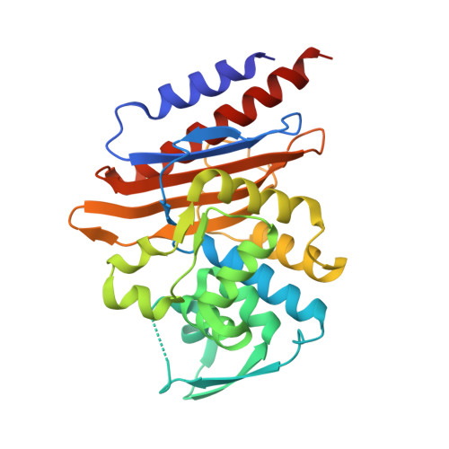
PER-2 belongs to a small (7 members to date) group of extended-spectrum β-lactamases. It has 88% amino acid identity with PER-1 and both display high catalytic efficiencies toward most β-lactams. In this study, we determined the X-ray structure of PER-2 at 2.20 Å and evaluated the possible role of several residues in the structure and activity toward β-lactams and mechanism-based inhibitors. PER-2 is defined by the presence of a singular trans bond between residues 166 to 167, which generates an inverted Ω loop, an expanded fold of this domain that results in a wide active site cavity that allows for efficient hydrolysis of antibiotics like the oxyimino-cephalosporins, and a series of exclusive interactions between residues not frequently involved in the stabilization of the active site in other class A β-lactamases. PER β-lactamases might be included within a cluster of evolutionarily related enzymes harboring the conserved residues Asp136 and Asn179. Other signature residues that define these enzymes seem to be Gln69, Arg220, Thr237, and probably Arg/Lys240A ("A" indicates an insertion according to Ambler's scheme for residue numbering in PER β-lactamases), with structurally important roles in the stabilization of the active site and proper orientation of catalytic water molecules, among others. We propose, supported by simulated models of PER-2 in combination with different β-lactams, the presence of a hydrogen-bond network connecting Ser70-Gln69-water-Thr237-Arg220 that might be important for the proper activity and inhibition of the enzyme. Therefore, we expect that mutations occurring in these positions will have impacts on the overall hydrolytic behavior.
- Laboratorio de Resistencia Bacteriana, Facultad de Farmacia y Bioquímica, Universidad de Buenos Aires, Buenos Aires, Argentina.
Organizational Affiliation: