4D2E | pdb_00004d2e

Crystal structure of an integral membrane kinase - v2.3


Experimental Data Snapshot

  • Method: X-RAY DIFFRACTION
  • Resolution: 2.28 Å
  • R-Value Free: 
    0.229 (Depositor), 0.234 (DCC) 
  • R-Value Work: 
    0.188 (Depositor), 0.194 (DCC) 
  • R-Value Observed: 
    0.190 (Depositor) 

Starting Model: experimental
View more details

wwPDB Validation 3D Report Full Report

Validation slider image for 4D2E

Ligand Structure Quality Assessment 


This is version 1.4 of the entry. See complete history

Literature

Cell-Free Expression and in Meso Crystallisation of an Integral Membrane Kinase for Structure Determination.

Boland, C.Li, D.Shah, S.T.A.Haberstock, S.Dotsch, V.Bernhard, F.Caffrey, M.

(2014) Cell Mol Life Sci 71: 4895

  • DOI: https://doi.org/10.1007/s00018-014-1655-7
  • Primary Citation Related Structures: 
    4BPD, 4D2E

  • PubMed Abstract: 

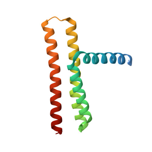
    Membrane proteins are key elements in cell physiology and drug targeting, but getting a high-resolution structure by crystallographic means is still enormously challenging. Novel strategies are in big demand to facilitate the structure determination process that will ultimately hasten the day when sequence information alone can provide a three-dimensional model. Cell-free or in vitro expression enables rapid access to large quantities of high-quality membrane proteins suitable for an array of applications. Despite its impressive efficiency, to date only two membrane proteins produced by the in vitro approach have yielded crystal structures. Here, we have analysed synergies of cell-free expression and crystallisation in lipid mesophases for generating an X-ray structure of the integral membrane enzyme diacylglycerol kinase to 2.28-Å resolution. The quality of cellular and cell-free-expressed kinase samples has been evaluated systematically by comparing (1) spectroscopic properties, (2) purity and oligomer formation, (3) lipid content and (4) functionality. DgkA is the first membrane enzyme crystallised based on cell-free expression. The study provides a basic standard for the crystallisation of cell-free-expressed membrane proteins and the methods detailed here should prove generally useful and contribute to accelerating the pace at which membrane protein structures are solved.


  • Organizational Affiliation
    • Membrane Structural and Functional Group, School of Medicine and School of Biochemistry and Immunology, Trinity College Dublin, Ireland.

Macromolecule Content 

  • Total Structure Weight: 91.35 kDa 
  • Atom Count: 5,344 
  • Modeled Residue Count: 597 
  • Deposited Residue Count: 780 
  • Unique protein chains: 1

Macromolecules

Find similar proteins by:|  3D Structure
Entity ID: 1
MoleculeChains  Sequence LengthOrganismDetailsImage
DIACYLGLYCEROL KINASE
A, B, C, D, E
A, B, C, D, E, F
130Escherichia coli K-12Mutation(s): 7 
EC: 2.7.1.107
Membrane Entity: Yes 
UniProt
Find proteins for P0ABN1 (Escherichia coli (strain K12))
Explore P0ABN1 
Go to UniProtKB:  P0ABN1
Entity Groups
Sequence Clusters30% Identity50% Identity70% Identity90% Identity95% Identity100% Identity
UniProt GroupP0ABN1
Sequence Annotations
Expand
Reference Sequence

Small Molecules

Ligands 5 Unique
IDChains Name / Formula / InChI Key2D Diagram3D Interactions
78M

Query on 78M


Download:Ideal Coordinates CCD File
G [auth A]
H [auth A]
I [auth A]
J [auth A]
O [auth C]
G [auth A],
H [auth A],
I [auth A],
J [auth A],
O [auth C],
P [auth C],
X [auth D]
(2S)-2,3-DIHYDROXYPROPYL(7Z)-PENTADEC-7-ENOATE
C18 H34 O4
BJMLBVHMHXYQFS-JJEJIETFSA-N
78N

Query on 78N


Download:Ideal Coordinates CCD File
AA [auth D]
BA [auth E]
K [auth A]
L [auth B]
M [auth B]
AA [auth D],
BA [auth E],
K [auth A],
L [auth B],
M [auth B],
N [auth B],
Q [auth C],
R [auth C],
S [auth C],
T [auth C],
Z [auth D]
(2R)-2,3-DIHYDROXYPROPYL(7Z)-PENTADEC-7-ENOATE
C18 H34 O4
BJMLBVHMHXYQFS-XZVRFQMRSA-N
FLC

Query on FLC


Download:Ideal Coordinates CCD File
V [auth D],
W [auth D]
CITRATE ANION
C6 H5 O7
KRKNYBCHXYNGOX-UHFFFAOYSA-K
ZN

Query on ZN


Download:Ideal Coordinates CCD File
Y [auth D]ZINC ION
Zn
PTFCDOFLOPIGGS-UHFFFAOYSA-N
NA

Query on NA


Download:Ideal Coordinates CCD File
U [auth D]SODIUM ION
Na
FKNQFGJONOIPTF-UHFFFAOYSA-N

Experimental Data & Validation

Experimental Data

  • Method: X-RAY DIFFRACTION
  • Resolution: 2.28 Å
  • R-Value Free:  0.229 (Depositor), 0.234 (DCC) 
  • R-Value Work:  0.188 (Depositor), 0.194 (DCC) 
  • R-Value Observed: 0.190 (Depositor) 
Space Group: P 21 21 21
Unit Cell:
Length ( Å )Angle ( ˚ )
a = 75.29α = 90
b = 91.82β = 90
c = 143.92γ = 90
Software Package:
Software NamePurpose
PHENIXrefinement
XDSdata reduction
XSCALEdata scaling
PHASERphasing

Structure Validation

View Full Validation Report


Ligand Structure Quality Assessment 


Entry History 

Deposition Data

Revision History  (Full details and data files)

  • Version 1.0: 2014-07-23
    Type: Initial release
  • Version 1.1: 2014-11-26
    Changes: Database references
  • Version 1.2: 2019-02-27
    Changes: Data collection, Experimental preparation, Other
  • Version 1.3: 2019-10-09
    Changes: Data collection, Derived calculations, Other
  • Version 1.4: 2023-12-20
    Changes: Data collection, Database references, Derived calculations, Refinement description