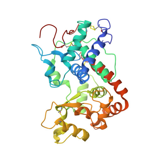
4CUO

Banyan peroxidase with glycosylation


Experimental Data Snapshot

  • Method: X-RAY DIFFRACTION
  • Resolution: 1.67 Å
  • R-Value Free: 0.180 
  • R-Value Work: 0.157 
  • R-Value Observed: 0.158 

wwPDB Validation   3D Report Full Report


Ligand Structure Quality Assessment 


This is version 3.0 of the entry. See complete history


Literature

Post-Translational Modification and Extended Glycosylation Pattern of a Plant Latex Peroxidase of Native Source Characterized by X-Ray Crystallography.

Palm, G.J.Sharma, A.Kumari, M.Panjikar, S.Albrecht, D.Jagannadham, M.V.Hinrichs, W.

(2014) FEBS J 281: 4319

  • DOI: https://doi.org/10.1111/febs.12900
  • Primary Citation of Related Structures:  
    4CUO

  • PubMed Abstract: 

    The crystal structure of banyan peroxidase purified from the latex of Ficus benghalensis has been solved at 1.67 Å resolution by single-wavelength anomalous diffraction phasing. The refined structure includes 306 amino acid residues, a heme and two calcium ions. The protein belongs to class III peroxidases and is the first one from plant latex. Extensive glycosylation was observed with N-linked glycans attached to seven asparagine residues. The enzyme is stable with respect to a wide pH range, temperature, chemical denaturants and organic solvents, probably as a result of its high glycosylation. An unexpected post-translational modification of Asp290 was identified as succinimide moiety. Kinetic parameters of banyan peroxidase have been determined using various hydrogen donor substrates and hydrogen peroxide.


  • Organizational Affiliation

    Institut für Biochemie, Universität Greifswald, Germany.


Macromolecules
Find similar proteins by:  (by identity cutoff)  |  3D Structure
Entity ID: 1
MoleculeChains Sequence LengthOrganismDetailsImage
BANYAN PEROXIDASE306Ficus benghalensisMutation(s): 0 
EC: 1.11.1.7
UniProt
Find proteins for A0A087WNH2 (Ficus benghalensis)
Explore A0A087WNH2 
Go to UniProtKB:  A0A087WNH2
Entity Groups  
Sequence Clusters30% Identity50% Identity70% Identity90% Identity95% Identity100% Identity
UniProt GroupA0A087WNH2
Sequence Annotations
Expand
  • Reference Sequence
Oligosaccharides

Help

Entity ID: 2
MoleculeChains Length2D Diagram Glycosylation3D Interactions
alpha-L-fucopyranose-(1-3)-2-acetamido-2-deoxy-beta-D-glucopyranose
B
2N-Glycosylation
Glycosylation Resources
GlyTouCan:  G80587NA
GlyCosmos:  G80587NA
GlyGen:  G80587NA
Entity ID: 3
MoleculeChains Length2D Diagram Glycosylation3D Interactions
alpha-D-xylopyranose-(1-2)-[alpha-D-mannopyranose-(1-3)][alpha-D-mannopyranose-(1-6)]beta-D-mannopyranose-(1-4)-2-acetamido-2-deoxy-beta-D-glucopyranose-(1-4)-[alpha-L-fucopyranose-(1-3)]2-acetamido-2-deoxy-beta-D-glucopyranose
C
7N-Glycosylation
Glycosylation Resources
GlyTouCan:  G98899DA
GlyCosmos:  G98899DA
GlyGen:  G98899DA
Entity ID: 4
MoleculeChains Length2D Diagram Glycosylation3D Interactions
alpha-L-fucopyranose-(1-3)-[2-acetamido-2-deoxy-beta-D-glucopyranose-(1-4)]2-acetamido-2-deoxy-beta-D-glucopyranose
D, E
3N-Glycosylation
Glycosylation Resources
GlyTouCan:  G11971MR
GlyCosmos:  G11971MR
GlyGen:  G11971MR
Small Molecules
Ligands 7 Unique
IDChains Name / Formula / InChI Key2D Diagram3D Interactions
HEM
Query on HEM

Download Ideal Coordinates CCD File 
F [auth A]PROTOPORPHYRIN IX CONTAINING FE
C34 H32 Fe N4 O4
KABFMIBPWCXCRK-RGGAHWMASA-L
NAG
Query on NAG

Download Ideal Coordinates CCD File 
G [auth A],
H [auth A],
I [auth A]
2-acetamido-2-deoxy-beta-D-glucopyranose
C8 H15 N O6
OVRNDRQMDRJTHS-FMDGEEDCSA-N
SO4
Query on SO4

Download Ideal Coordinates CCD File 
P [auth A],
Q [auth A],
R [auth A]
SULFATE ION
O4 S
QAOWNCQODCNURD-UHFFFAOYSA-L
CO3
Query on CO3

Download Ideal Coordinates CCD File 
S [auth A],
T [auth A],
U [auth A],
V [auth A]
CARBONATE ION
C O3
BVKZGUZCCUSVTD-UHFFFAOYSA-L
CA
Query on CA

Download Ideal Coordinates CCD File 
J [auth A],
K [auth A]
CALCIUM ION
Ca
BHPQYMZQTOCNFJ-UHFFFAOYSA-N
CL
Query on CL

Download Ideal Coordinates CCD File 
M [auth A],
N [auth A],
O [auth A]
CHLORIDE ION
Cl
VEXZGXHMUGYJMC-UHFFFAOYSA-M
NA
Query on NA

Download Ideal Coordinates CCD File 
L [auth A]SODIUM ION
Na
FKNQFGJONOIPTF-UHFFFAOYSA-N
Modified Residues  1 Unique
IDChains TypeFormula2D DiagramParent
SNN
Query on SNN
A
L-PEPTIDE LINKINGC4 H6 N2 O2ASN
Experimental Data & Validation

Experimental Data

  • Method: X-RAY DIFFRACTION
  • Resolution: 1.67 Å
  • R-Value Free: 0.180 
  • R-Value Work: 0.157 
  • R-Value Observed: 0.158 
  • Space Group: P 32 2 1
Unit Cell:
Length ( Å )Angle ( ˚ )
a = 73.115α = 90
b = 73.115β = 90
c = 164.596γ = 120
Software Package:
Software NamePurpose
REFMACrefinement
XDSdata reduction
XDSdata scaling
Auto-Rickshawphasing

Structure Validation

View Full Validation Report


Ligand Structure Quality Assessment 


Entry History 

Deposition Data

Revision History  (Full details and data files)

  • Version 1.0: 2014-07-23
    Type: Initial release
  • Version 1.1: 2014-10-01
    Changes: Database references
  • Version 1.2: 2019-07-17
    Changes: Data collection, Derived calculations
  • Version 2.0: 2020-07-29
    Type: Remediation
    Reason: Carbohydrate remediation
    Changes: Advisory, Atomic model, Data collection, Derived calculations, Other, Structure summary
  • Version 3.0: 2023-11-15
    Changes: Atomic model, Data collection, Database references, Derived calculations, Structure summary