4CP9 | pdb_00004cp9

Crystal structure OF lecA lectin complexed with a divalent galactoside at 1.65 angstrom


Experimental Data Snapshot

  • Method: X-RAY DIFFRACTION
  • Resolution: 1.65 Å
  • R-Value Free: 
    0.184 (Depositor), 0.193 (DCC) 
  • R-Value Work: 
    0.155 (Depositor), 0.168 (DCC) 
  • R-Value Observed: 
    0.157 (Depositor) 

Starting Model: experimental
View more details

wwPDB Validation 3D Report Full Report

Validation slider image for 4CP9

Ligand Structure Quality Assessment 


This is version 1.4 of the entry. See complete history

Literature

A Leca Ligand Identified from a Galactoside-Conjugate Array Inhibits Host Cell Invasion by Pseudomonas Aeruginosa.

Novoa, A.Eierhoff, T.Topin, J.Varrot, A.Barluenga, S.Imberty, A.Romer, W.Winssinger, N.

(2014) Angew Chem Int Ed Engl 53: 8885

  • DOI: https://doi.org/10.1002/anie.201402831
  • Primary Citation Related Structures: 
    4CP9, 4CPB

  • PubMed Abstract: 

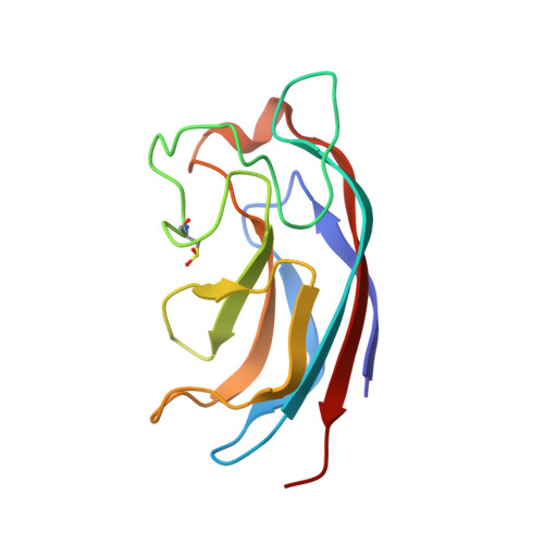
    Lectin LecA is a virulence factor of Pseudomonas aeruginosa involved in lung injury, mortality, and cellular invasion. Ligands competing with human glycoconjugates for LecA binding are thus promising candidates to counteract P. aeruginosa infections. We have identified a novel divalent ligand from a focused galactoside(Gal)-conjugate array which binds to LecA with very high affinity (Kd = 82 nM). Crystal structures of LecA complexed with the ligand together with modeling studies confirmed its ability to chelate two binding sites of LecA. The ligand lowers cellular invasiveness of P. aeruginosa up to 90 % when applied in the range of 0.05-5 μM. Hence, this ligand might lead to the development of drugs against P. aeruginosa infection.


  • Organizational Affiliation
    • Department of Organic Chemistry, University of Geneva (Switzerland).

Macromolecule Content 

  • Total Structure Weight: 55.39 kDa 
  • Atom Count: 4,294 
  • Modeled Residue Count: 484 
  • Deposited Residue Count: 484 
  • Unique protein chains: 2

Macromolecules

Find similar proteins by:|  3D Structure
Entity ID: 1
MoleculeChains  Sequence LengthOrganismDetailsImage
PA-I GALACTOPHILIC LECTIN121Pseudomonas aeruginosa PAO1Mutation(s): 0 
UniProt
Find proteins for Q05097 (Pseudomonas aeruginosa (strain ATCC 15692 / DSM 22644 / CIP 104116 / JCM 14847 / LMG 12228 / 1C / PRS 101 / PAO1))
Explore Q05097 
Go to UniProtKB:  Q05097
Entity Groups
Sequence Clusters30% Identity50% Identity70% Identity90% Identity95% Identity100% Identity
UniProt GroupQ05097
Sequence Annotations
Expand
Reference Sequence
Find similar proteins by:|  3D Structure
Entity ID: 2
MoleculeChains  Sequence LengthOrganismDetailsImage
PA-I GALACTOPHILIC LECTIN
B, C, D
121Pseudomonas aeruginosa PAO1Mutation(s): 0 
UniProt
Find proteins for Q05097 (Pseudomonas aeruginosa (strain ATCC 15692 / DSM 22644 / CIP 104116 / JCM 14847 / LMG 12228 / 1C / PRS 101 / PAO1))
Explore Q05097 
Go to UniProtKB:  Q05097
Entity Groups
Sequence Clusters30% Identity50% Identity70% Identity90% Identity95% Identity100% Identity
UniProt GroupQ05097
Sequence Annotations
Expand
Reference Sequence

Small Molecules

Ligands 6 Unique
IDChains Name / Formula / InChI Key2D Diagram3D Interactions
CN8

Query on CN8


Download:Ideal Coordinates CCD File
G [auth A],
L [auth B],
R [auth C],
V [auth D]
(4S)-N-ethyl-4-{[N-methyl-3-(1-{2-[(4-sulfanylbenzoyl)amino]ethyl}-1H-1,2,3-triazol-4-yl)-L-alanyl]amino}-L-prolinamide
C22 H32 N8 O3 S
VTNMGGUGQYETGZ-SNRMKQJTSA-N
1PE

Query on 1PE


Download:Ideal Coordinates CCD File
H [auth A],
M [auth B],
S [auth C],
X [auth D]
PENTAETHYLENE GLYCOL
C10 H22 O6
JLFNLZLINWHATN-UHFFFAOYSA-N
GAL

Query on GAL


Download:Ideal Coordinates CCD File
F [auth A],
K [auth B],
Q [auth C],
U [auth D]
beta-D-galactopyranose
C6 H12 O6
WQZGKKKJIJFFOK-FPRJBGLDSA-N
SO4

Query on SO4


Download:Ideal Coordinates CCD File
I [auth A],
W [auth D]
SULFATE ION
O4 S
QAOWNCQODCNURD-UHFFFAOYSA-L
EDO

Query on EDO


Download:Ideal Coordinates CCD File
N [auth B],
O [auth B]
1,2-ETHANEDIOL
C2 H6 O2
LYCAIKOWRPUZTN-UHFFFAOYSA-N
CA

Query on CA


Download:Ideal Coordinates CCD File
E [auth A],
J [auth B],
P [auth C],
T [auth D]
CALCIUM ION
Ca
BHPQYMZQTOCNFJ-UHFFFAOYSA-N
Modified Residues  2 Unique
IDChains TypeFormula2D DiagramParent
OCS
Query on OCS
A
L-PEPTIDE LINKINGC3 H7 N O5 SCYS
TRO
Query on TRO
A
L-PEPTIDE LINKINGC11 H12 N2 O3TRP
Binding Affinity Annotations 
IDSourceBinding Affinity
GAL BindingDB:  4CP9 Kd: 8.75e+4 (nM) from 1 assay(s)

Experimental Data & Validation

Experimental Data

  • Method: X-RAY DIFFRACTION
  • Resolution: 1.65 Å
  • R-Value Free:  0.184 (Depositor), 0.193 (DCC) 
  • R-Value Work:  0.155 (Depositor), 0.168 (DCC) 
  • R-Value Observed: 0.157 (Depositor) 
Space Group: P 21 21 21
Unit Cell:
Length ( Å )Angle ( ˚ )
a = 50.876α = 90
b = 66.259β = 90
c = 159.427γ = 90
Software Package:
Software NamePurpose
REFMACrefinement
XDSdata reduction
Aimlessdata scaling
PHASERphasing

Structure Validation

View Full Validation Report


Ligand Structure Quality Assessment 


Entry History 

Deposition Data

Revision History  (Full details and data files)

  • Version 1.0: 2014-10-08
    Type: Initial release
  • Version 1.1: 2015-04-22
    Changes: Derived calculations, Non-polymer description, Other
  • Version 1.2: 2016-12-28
    Changes: Structure summary
  • Version 1.3: 2020-07-29
    Type: Remediation
    Reason: Carbohydrate remediation
    Changes: Data collection, Derived calculations, Other, Structure summary
  • Version 1.4: 2023-12-20
    Changes: Data collection, Database references, Refinement description, Structure summary