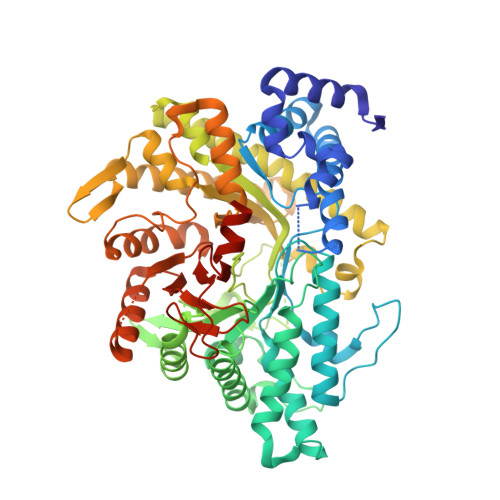
The Crystal Structure of Thermotoga Maritima Class III Ribonucleotide Reductase Lacks a Radical Cysteine Pre-Positioned in the Active Site.
Aurelius, O., Johansson, R., Bagenholm, V., Beck, T., Balhuizen, A., Lundin, D., Sjoberg, B.M., Mulliez, E., Logan, D.T.(2015) PLoS One 10: 01281
- PubMed: 26147435 Search on PubMedSearch on PubMed Central
- DOI: https://doi.org/10.1371/journal.pone.0128199
- Primary Citation Related Structures:
4COI, 4COJ, 4COL, 4COM, 4CON - PubMed Abstract:
Ribonucleotide reductases (RNRs) catalyze the reduction of ribonucleotides to deoxyribonucleotides, the building blocks for DNA synthesis, and are found in all but a few organisms. RNRs use radical chemistry to catalyze the reduction reaction. Despite RNR having evolved several mechanisms for generation of different kinds of essential radicals across a large evolutionary time frame, this initial radical is normally always channelled to a strictly conserved cysteine residue directly adjacent to the substrate for initiation of substrate reduction, and this cysteine has been found in the structures of all RNRs solved to date. We present the crystal structure of an anaerobic RNR from the extreme thermophile Thermotoga maritima (tmNrdD), alone and in several complexes, including with the allosteric effector dATP and its cognate substrate CTP. In the crystal structure of the enzyme as purified, tmNrdD lacks a cysteine for radical transfer to the substrate pre-positioned in the active site. Nevertheless activity assays using anaerobic cell extracts from T. maritima demonstrate that the class III RNR is enzymatically active. Other genetic and microbiological evidence is summarized indicating that the enzyme is important for T. maritima. Mutation of either of two cysteine residues in a disordered loop far from the active site results in inactive enzyme. We discuss the possible mechanisms for radical initiation of substrate reduction given the collected evidence from the crystal structure, our activity assays and other published work. Taken together, the results suggest either that initiation of substrate reduction may involve unprecedented conformational changes in the enzyme to bring one of these cysteine residues to the expected position, or that alternative routes for initiation of the RNR reduction reaction may exist. Finally, we present a phylogenetic analysis showing that the structure of tmNrdD is representative of a new RNR subclass IIIh, present in all Thermotoga species plus a wider group of bacteria from the distantly related phyla Firmicutes, Bacteroidetes and Proteobacteria.
- Dept. of Biochemistry & Structural Biology, Lund University, Box 124, S-221 00 Lund, Sweden.
Organizational Affiliation: