4CI5 | pdb_00004ci5

Structural basis for GL479 a dual Peroxisome Proliferator-Activated Receptor gamma agonist


Experimental Data Snapshot

  • Method: X-RAY DIFFRACTION
  • Resolution: 1.77 Å
  • R-Value Free: 
    0.212 (Depositor), 0.215 (DCC) 
  • R-Value Work: 
    0.175 (Depositor), 0.181 (DCC) 
  • R-Value Observed: 
    0.177 (Depositor) 

Starting Model: experimental
View more details

wwPDB Validation 3D Report Full Report

Validation slider image for 4CI5

Ligand Structure Quality Assessment 


This is version 1.4 of the entry. See complete history

Literature

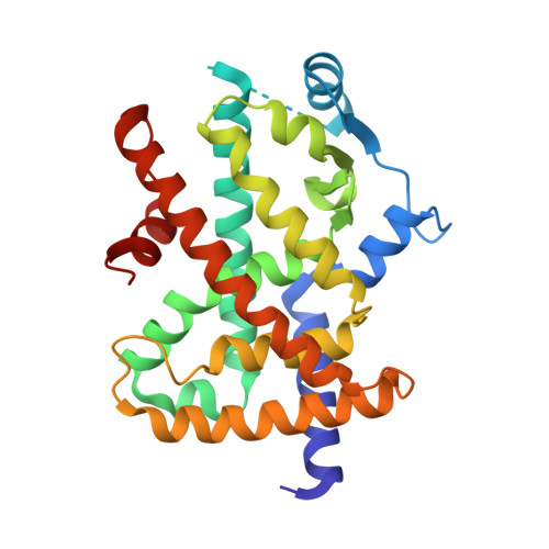
Different binding and recognition modes of GL479, a dual agonist of Peroxisome Proliferator-Activated Receptor alpha / gamma.

dos Santos, J.C.Bernardes, A.Giampietro, L.Ammazzalorso, A.De Filippis, B.Amoroso, R.Polikarpov, I.

(2015) J Struct Biol 191: 332-340

  • DOI: https://doi.org/10.1016/j.jsb.2015.07.006
  • Primary Citation Related Structures: 
    4CI4, 4CI5

  • PubMed Abstract: 

    Peroxisome Proliferator-Activated Receptors (PPARs) are ligand-dependent transcription factors that control various functions in human organism, including the control of glucose and lipid metabolism. PPARγ is a target of TZD agonists, clinically used to improve insulin sensitivity whereas fibrates, PPARα ligands, lower serum triglyceride levels. We report here the structural studies of GL479, a synthetic dual PPARα/γ agonist, designed by a combination of clofibric acid skeleton and a phenyldiazenyl moiety, as bioisosteric replacement of stilbene group, in complex with both PPARα and PPARγ receptors. GL479 was previously reported as a partial agonist of PPARγ and a full agonist of PPARα with high affinity for both PPARs. Our structural studies reveal different binding modes of GL479 to PPARα and PPARγ, which may explain the distinct activation behaviors observed for each receptor. In both cases the ligand interacts with a Tyr located at helix 12 (H12), resulting in the receptor active conformation. In the complex with PPARα, GL479 occupies the same region of the ligand-binding pocket (LBP) observed for other full agonists, whereas GL479 bound to PPARγ displays a new binding mode. Our results indicate a novel region of PPARs LBP that may be explored for the design of partial agonists as well dual PPARα/γ agonists that combine, simultaneously, the therapeutic effects of the treatment of insulin resistance and dyslipidemia.


  • Organizational Affiliation
    • Grupo de Biotecnologia Molecular, Instituto de Física de São Carlos, Universidade de São Paulo, São Carlos, SP 13566-590, Brazil.

Macromolecule Content 

  • Total Structure Weight: 63 kDa 
  • Atom Count: 4,686 
  • Modeled Residue Count: 520 
  • Deposited Residue Count: 544 
  • Unique protein chains: 1

Macromolecules

Find similar proteins by:|  3D Structure
Entity ID: 1
MoleculeChains  Sequence LengthOrganismDetailsImage
PEROXISOME PROLIFERATOR-ACTIVATED RECEPTOR GAMMA
A, B
272Homo sapiensMutation(s): 0 
UniProt & NIH Common Fund Data Resources
Find proteins for P37231 (Homo sapiens)
Explore P37231 
Go to UniProtKB:  P37231
PHAROS:  P37231
GTEx:  ENSG00000132170 
Entity Groups
Sequence Clusters30% Identity50% Identity70% Identity90% Identity95% Identity100% Identity
UniProt GroupP37231
Sequence Annotations
Expand
Reference Sequence

Small Molecules

Ligands 1 Unique
IDChains Name / Formula / InChI Key2D Diagram3D Interactions
Y1N

Query on Y1N


Download:Ideal Coordinates CCD File
C [auth A],
D [auth B]
2-methyl-2-[4-[2-[4-[(E)-phenyldiazenyl]phenoxy]ethyl]phenoxy]propanoic acid
C24 H24 N2 O4
HCISJKNVUOHNQX-OCEACIFDSA-N

Experimental Data & Validation

Experimental Data

  • Method: X-RAY DIFFRACTION
  • Resolution: 1.77 Å
  • R-Value Free:  0.212 (Depositor), 0.215 (DCC) 
  • R-Value Work:  0.175 (Depositor), 0.181 (DCC) 
  • R-Value Observed: 0.177 (Depositor) 
Space Group: C 1 2 1
Unit Cell:
Length ( Å )Angle ( ˚ )
a = 88.488α = 90
b = 64.247β = 102.59
c = 118.781γ = 90
Software Package:
Software NamePurpose
PHENIXrefinement
XDSdata reduction
SCALAdata scaling
PHASERphasing

Structure Validation

View Full Validation Report


Ligand Structure Quality Assessment 


Entry History 

Deposition Data

Revision History  (Full details and data files)

  • Version 1.0: 2014-12-24
    Type: Initial release
  • Version 1.1: 2015-08-05
    Changes: Database references
  • Version 1.2: 2015-09-09
    Changes: Database references
  • Version 1.3: 2019-01-16
    Changes: Data collection, Database references
  • Version 1.4: 2023-12-20
    Changes: Data collection, Database references, Derived calculations, Other, Refinement description