4CDK

Structure of ZNRF3-RSPO1


Experimental Data Snapshot

  • Method: X-RAY DIFFRACTION
  • Resolution: 2.80 Å
  • R-Value Free: 0.246 
  • R-Value Work: 0.218 
  • R-Value Observed: 0.219 

wwPDB Validation   3D Report Full Report


This is version 1.1 of the entry. See complete history


Literature

Structures of Wnt-Antagonist Znrf3 and its Complex with R-Spondin 1 and Implications for Signaling.

Peng, W.C.De Lau, W.Madoori, P.K.Forneris, F.Granneman, J.C.M.Clevers, H.Gros, P.

(2013) PLoS One 8: 83110

  • DOI: https://doi.org/10.1371/journal.pone.0083110
  • Primary Citation of Related Structures:  
    4CDJ, 4CDK

  • PubMed Abstract: 

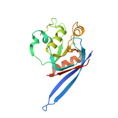
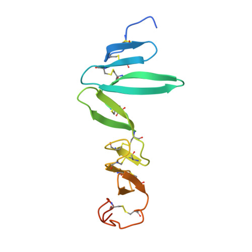
    Zinc RING finger 3 (ZNRF3) and its homolog RING finger 43 (RNF43) antagonize Wnt signaling in adult stem cells by ubiquitinating Frizzled receptors (FZD), which leads to endocytosis of the Wnt receptor. Conversely, binding of ZNRF3/RNF43 to LGR4-6 - R-spondin blocks Frizzled ubiquitination and enhances Wnt signaling. Here, we present crystal structures of the ZNRF3 ectodomain and its complex with R-spondin 1 (RSPO1). ZNRF3 binds RSPO1 and LGR5-RSPO1 with micromolar affinity via RSPO1 furin-like 1 (Fu1) domain. Anonychia-related mutations in RSPO4 support the importance of the observed interface. The ZNRF3-RSPO1 structure resembles that of LGR5-RSPO1-RNF43, though Fu2 of RSPO1 is variably oriented. The ZNRF3-binding site overlaps with trans-interactions observed in 2:2 LGR5-RSPO1 complexes, thus binding of ZNRF3/RNF43 would disrupt such an arrangement. Sequence conservation suggests a single ligand-binding site on ZNRF3, consistent with the proposed competing binding role of ZNRF3/RNF43 in Wnt signaling.


  • Organizational Affiliation

    Crystal and Structural Chemistry, Bijvoet Center for Biomolecular Research, Department of Chemistry, Faculty of Science, Utrecht University, Utrecht, The Netherlands.


Macromolecules
Find similar proteins by:  (by identity cutoff)  |  3D Structure
Entity ID: 1
MoleculeChains Sequence LengthOrganismDetailsImage
E3 UBIQUITIN-PROTEIN LIGASE ZNRF3
A, B, C, D
164Mus musculusMutation(s): 0 
UniProt
Find proteins for Q5SSZ7 (Mus musculus)
Explore Q5SSZ7 
Go to UniProtKB:  Q5SSZ7
Entity Groups  
Sequence Clusters30% Identity50% Identity70% Identity90% Identity95% Identity100% Identity
UniProt GroupQ5SSZ7
Sequence Annotations
Expand
  • Reference Sequence
Find similar proteins by:  (by identity cutoff)  |  3D Structure
Entity ID: 2
MoleculeChains Sequence LengthOrganismDetailsImage
R-SPONDIN-1
E, F, G, H
126Homo sapiensMutation(s): 0 
UniProt & NIH Common Fund Data Resources
Find proteins for Q2MKA7 (Homo sapiens)
Explore Q2MKA7 
Go to UniProtKB:  Q2MKA7
PHAROS:  Q2MKA7
GTEx:  ENSG00000169218 
Entity Groups  
Sequence Clusters30% Identity50% Identity70% Identity90% Identity95% Identity100% Identity
UniProt GroupQ2MKA7
Sequence Annotations
Expand
  • Reference Sequence
Experimental Data & Validation

Experimental Data

  • Method: X-RAY DIFFRACTION
  • Resolution: 2.80 Å
  • R-Value Free: 0.246 
  • R-Value Work: 0.218 
  • R-Value Observed: 0.219 
  • Space Group: P 1
Unit Cell:
Length ( Å )Angle ( ˚ )
a = 51.7α = 66.27
b = 80.16β = 81.36
c = 82.98γ = 80.66
Software Package:
Software NamePurpose
PHENIXrefinement

Structure Validation

View Full Validation Report


Entry History 

Deposition Data

Revision History  (Full details and data files)

  • Version 1.0: 2014-01-08
    Type: Initial release
  • Version 1.1: 2019-04-03
    Changes: Data collection, Other, Source and taxonomy