4C39 | pdb_00004c39

Structure of rat neuronal nitric oxide synthase heme domain in complex with 6-((((3S, 5R)-5-(((6-amino-4-methylpyridin-2-yl)methoxy) methyl)pyrrolidin-3-yl)oxy)methyl)-4-methylpyridin-2-amine


Experimental Data Snapshot

  • Method: X-RAY DIFFRACTION
  • Resolution: 1.98 Å
  • R-Value Free: 
    0.228 (Depositor), 0.228 (DCC) 
  • R-Value Work: 
    0.181 (Depositor), 0.180 (DCC) 
  • R-Value Observed: 
    0.183 (Depositor) 

wwPDB Validation 3D Report Full Report

Validation slider image for 4C39

Ligand Structure Quality Assessment 


This is version 1.1 of the entry. See complete history

Literature

An Accessible Chiral Linker to Enhance Potency and Selectivity of Neuronal Nitric Oxide Synthase Inhibitors.

Jing, Q.Li, H.Roman, L.J.Martasek, P.Poulos, T.L.Silverman, R.B.

(2014) ACS Med Chem Lett 5: 56

  • DOI: https://doi.org/10.1021/ml400381s
  • Primary Citation Related Structures: 
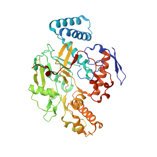
    4C39, 4C3A

  • PubMed Abstract: 

    The three important mammalian isozymes of nitric oxide synthase (NOS) are neuronal NOS (nNOS), endothelial NOS (eNOS), and inducible NOS (iNOS). Inhibitors of nNOS show promise as treatments for neurodegenerative diseases. Eight easily-synthesized compounds containing either one ( 20a,b ) or two ( 9a-d; 15a,b ) 2-amino-4-methylpyridine groups with a chiral pyrrolidine linker were designed as selective nNOS inhibitors. Inhibitor 9c is the best of these compounds, having a potency of 9.7 nM and dual selectivity of 693 and 295 against eNOS and iNOS, respectively. Crystal structures of nNOS complexed with either 9a or 9c show a double-headed binding mode, where each 2-aminopyridine head group interacts with either a nNOS active site Glu residue or a heme propionate. In addition, the pyrrolidine nitrogen of 9c contributes additional hydrogen bonds to the heme propionate, resulting in a unique binding orientation. In contrast, the lack of hydrogen bonds from the pyrrolidine of 9a to the heme propionate allows the inhibitor to adopt two different binding orientations. Both 9a and 9c bind to eNOS in a single-headed mode, which is the structural basis for the isozyme selectivity.


  • Organizational Affiliation
    • Department of Chemistry, Department of Molecular Biosciences, Chemistry of Life Processes Institute, Center for Molecular Innovation and Drug Discovery, Northwestern University, Evanston, Illinois 60208-3113, USA.

Macromolecule Content 

  • Total Structure Weight: 100.24 kDa 
  • Atom Count: 7,179 
  • Modeled Residue Count: 819 
  • Deposited Residue Count: 844 
  • Unique protein chains: 1

Macromolecules

Find similar proteins by:|  3D Structure
Entity ID: 1
MoleculeChains  Sequence LengthOrganismDetailsImage
NITRIC OXIDE SYNTHASE, BRAIN
A, B
422Rattus norvegicusMutation(s): 0 
EC: 1.14.13.39
UniProt
Find proteins for P29476 (Rattus norvegicus)
Explore P29476 
Go to UniProtKB:  P29476
Entity Groups
Sequence Clusters30% Identity50% Identity70% Identity90% Identity95% Identity100% Identity
UniProt GroupP29476
Sequence Annotations
Expand
Reference Sequence

Small Molecules

Ligands 5 Unique
IDChains Name / Formula / InChI Key2D Diagram3D Interactions
HEM

Query on HEM


Download:Ideal Coordinates CCD File
C [auth A],
H [auth B]
PROTOPORPHYRIN IX CONTAINING FE
C34 H32 Fe N4 O4
KABFMIBPWCXCRK-RGGAHWMASA-L
Q16

Query on Q16


Download:Ideal Coordinates CCD File
E [auth A],
J [auth B]
6-((((3S, 5R)-5-(((6-amino-4-methylpyridin-2-yl)methoxy)methyl)pyrrolidin-3-yl)oxy)methyl)-4-methylpyridin-2-amine
C19 H27 N5 O2
NUKSCYDKRQJOFO-PBHICJAKSA-N
H4B

Query on H4B


Download:Ideal Coordinates CCD File
D [auth A],
I [auth B]
5,6,7,8-TETRAHYDROBIOPTERIN
C9 H15 N5 O3
FNKQXYHWGSIFBK-RPDRRWSUSA-N
ZN

Query on ZN


Download:Ideal Coordinates CCD File
G [auth A]ZINC ION
Zn
PTFCDOFLOPIGGS-UHFFFAOYSA-N
ACT

Query on ACT


Download:Ideal Coordinates CCD File
F [auth A],
K [auth B]
ACETATE ION
C2 H3 O2
QTBSBXVTEAMEQO-UHFFFAOYSA-M
Binding Affinity Annotations 
IDSourceBinding Affinity
Q16 BindingDB:  4C39 Ki: 9.7 (nM) from 1 assay(s)

Experimental Data & Validation

Experimental Data

  • Method: X-RAY DIFFRACTION
  • Resolution: 1.98 Å
  • R-Value Free:  0.228 (Depositor), 0.228 (DCC) 
  • R-Value Work:  0.181 (Depositor), 0.180 (DCC) 
  • R-Value Observed: 0.183 (Depositor) 
Space Group: P 21 21 21
Unit Cell:
Length ( Å )Angle ( ˚ )
a = 51.929α = 90
b = 110.87β = 90
c = 164.439γ = 90
Software Package:
Software NamePurpose
REFMACrefinement
HKL-2000data reduction
SCALEPACKdata scaling
REFMACphasing

Structure Validation

View Full Validation Report


Ligand Structure Quality Assessment 


Entry History 

Deposition Data

Revision History  (Full details and data files)

  • Version 1.0: 2014-04-02
    Type: Initial release
  • Version 1.1: 2024-05-08
    Changes: Data collection, Database references, Derived calculations, Other