Mechanistic Implications for the Chorismatase Fkbo Based on the Crystal Structure.
Juneja, P., Hubrich, F., Diederichs, K., Welte, W., Andexer, J.N.(2014) J Mol Biology 426: 105
- PubMed: 24036425 Search on PubMed
- DOI: https://doi.org/10.1016/j.jmb.2013.09.006
- Primary Citation Related Structures:
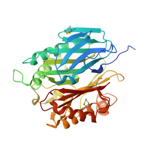
4BPS - PubMed Abstract:
Chorismate-converting enzymes are involved in many biosynthetic pathways leading to natural products and can often be used as tools for the synthesis of chemical building blocks. Chorismatases such as FkbO from Streptomyces species catalyse the hydrolysis of chorismate yielding (dihydro)benzoic acid derivatives. In contrast to many other chorismate-converting enzymes, the structure and catalytic mechanism of a chorismatase had not been previously elucidated. Here we present the crystal structure of the chorismatase FkbO in complex with a competitive inhibitor at 1.08Å resolution. FkbO is a monomer in solution and exhibits pseudo-3-fold symmetry; the structure of the individual domains indicates a possible connection to the trimeric RidA/YjgF family and related enzymes. The co-crystallised inhibitor led to the identification of FkbO's active site in the cleft between the central and the C-terminal domains. A mechanism for FkbO is proposed based on both interactions between the inhibitor and the surrounding amino acids and an FkbO structure with chorismate modelled in the active site. We suggest that the methylene group of the chorismate enol ether takes up a proton from an active-site glutamic acid residue, thereby initiating chorismate hydrolysis. A similar chemistry has been described for isochorismatases, albeit implemented in an entirely different protein scaffold. This reaction model is supported by kinetic data from active-site variants of FkbO derived by site-directed mutagenesis.
- Department of Biology, University of Konstanz, D-78457 Konstanz, Germany.
Organizational Affiliation: