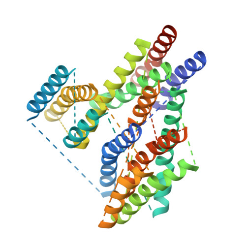
Structure and Substrate-Induced Conformational Changes of the Secondary Citrate/Sodium Symporter Cits Revealed by Electron Crystallography.
Kebbel, F., Kurz, M., Arheit, M., Grutter, M.G., Stahlberg, H.(2013) Structure 21: 1243
- PubMed: 23810698 Search on PubMed
- DOI: https://doi.org/10.1016/j.str.2013.05.011
- Primary Citation Related Structures:
4BPQ - PubMed Abstract:
The secondary Na+/citrate symporter CitS of Klebsiella pneumoniae is the best-characterized member of the 2-hydroxycarboxylate transporter family. The recent projection structure gave insight into its overall structural organization. Here, we present the three-dimensional map of dimeric CitS obtained with electron crystallography. Each monomer has 13 a-helical transmembrane segments; six are organized in a distal helix cluster and seven in the central dimer interface domain. Based on structural analyses and comparison to VcINDY, we propose a molecular model for CitS, assign the helices, and demonstrate the internal structural symmetry. We also present projections of CitS in several conformational states induced by the presence and absence of sodium and citrate as substrates. Citrate binding induces a defined movement of a helices within the distal helical cluster. Based on this, we propose a substrate translocation site and conformational changes that are in agreement with the transport model of ‘‘alternating access’’.
- Center for Cellular Imaging and NanoAnalytics (C-CINA), Biozentrum, University Basel, Mattenstrasse 26, CH-4058 Basel, Switzerland.
Organizational Affiliation: