Structural Enzymology of Cellvibrio Japonicus Agd31B Reveals Alpha-Transglucosylase Activity in Glycoside Hydrolase Family 31
Larsbrink, J., Izumi, A., Hemsworth, G.R., Davies, G.J., Brumer, H.(2012) J Biol Chem 287: 43288
- PubMed: 23132856 Search on PubMedSearch on PubMed Central
- DOI: https://doi.org/10.1074/jbc.M112.416511
- Primary Citation Related Structures:
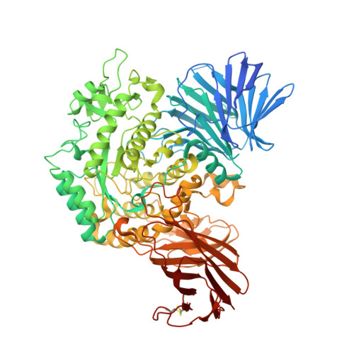
4B9Y, 4B9Z, 4BA0 - PubMed Abstract:
The metabolism of the storage polysaccharides glycogen and starch is of vital importance to organisms from all domains of life. In bacteria, utilization of these α-glucans requires the concerted action of a variety of enzymes, including glycoside hydrolases, glycoside phosphorylases, and transglycosylases. In particular, transglycosylases from glycoside hydrolase family 13 (GH13) and GH77 play well established roles in α-glucan side chain (de)branching, regulation of oligo- and polysaccharide chain length, and formation of cyclic dextrans. Here, we present the biochemical and tertiary structural characterization of a new type of bacterial 1,4-α-glucan 4-α-glucosyltransferase from GH31. Distinct from 1,4-α-glucan 6-α-glucosyltransferases (EC 2.4.1.24) and 4-α-glucanotransferases (EC 2.4.1.25), this enzyme strictly transferred one glucosyl residue from α(1→4)-glucans in disproportionation reactions. Substrate hydrolysis was undetectable for a series of malto-oligosaccharides except maltose for which transglycosylation nonetheless dominated across a range of substrate concentrations. Crystallographic analysis of the enzyme in free, acarbose-complexed, and trapped 5-fluoro-β-glucosyl-enzyme intermediate forms revealed extended substrate interactions across one negative and up to three positive subsites, thus providing structural rationalization for the unique, single monosaccharide transferase activity of the enzyme.
- Division of Glycoscience, School of Biotechnology, Royal Institute of Technology, AlbaNova University Centre, 106 91 Stockholm, Sweden.
Organizational Affiliation: