4B5P

Crystal structure of human alpha tubulin acetyltransferase catalytic domain Q58A variant


Experimental Data Snapshot

  • Method: X-RAY DIFFRACTION
  • Resolution: 1.60 Å
  • R-Value Free: 0.222 
  • R-Value Work: 0.185 
  • R-Value Observed: 0.186 

wwPDB Validation   3D Report Full Report


Ligand Structure Quality Assessment 


This is version 1.3 of the entry. See complete history


Literature

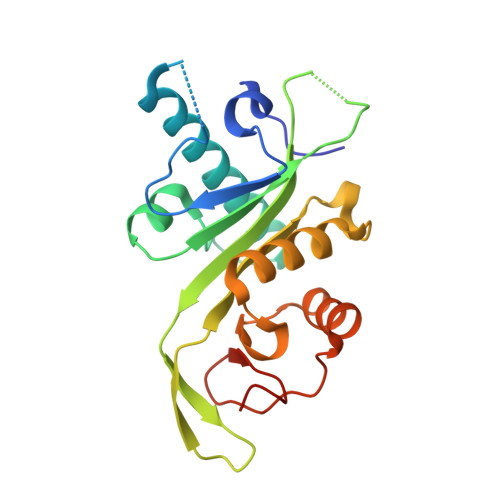
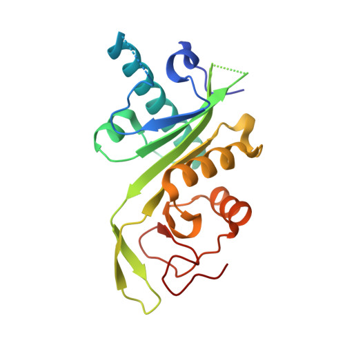
Atomic Resolution Structure of Human Alpha-Tubulin Acetyltransferase Bound to Acetyl-Coa.

Taschner, M.Vetter, M.Lorentzen, E.

(2012) Proc Natl Acad Sci U S A 109: 19649

  • DOI: https://doi.org/10.1073/pnas.1209343109
  • Primary Citation of Related Structures:  
    4B5O, 4B5P

  • PubMed Abstract: 

    Acetylation of lysine residues is an important posttranslational modification found in all domains of life. α-Tubulin is specifically acetylated on lysine 40, a modification that serves to stabilize microtubules of axons and cilia. Whereas histone acetyltransferases have been extensively studied, there is no structural and mechanistic information available on α-tubulin acetyltransferases. Here, we present the structure of the human α-tubulin acetyltransferase catalytic domain bound to its cosubstrate acetyl-CoA at 1.05 Å resolution. Compared with other lysine acetyltransferases of known structure, α-tubulin acetyltransferase displays a relatively well-conserved cosubstrate binding pocket but is unique in its active site and putative α-tubulin binding site. Using acetylation assays with structure-guided mutants, we map residues important for acetyl-CoA binding, substrate binding, and catalysis. This analysis reveals a basic patch implicated in substrate binding and a conserved glutamine residue required for catalysis, demonstrating that the family of α-tubulin acetyltransferases uses a reaction mechanism different from other lysine acetyltransferases characterized to date.


  • Organizational Affiliation

    Department of Structural Cell Biology, Max Planck Institute of Biochemistry, D-82152 Martinsried, Germany.


Macromolecules
Find similar proteins by:  (by identity cutoff)  |  3D Structure
Entity ID: 1
MoleculeChains Sequence LengthOrganismDetailsImage
ALPHA-TUBULIN N-ACETYLTRANSFERASE200Homo sapiensMutation(s): 1 
EC: 2.3.1.108
UniProt & NIH Common Fund Data Resources
Find proteins for Q5SQI0 (Homo sapiens)
Explore Q5SQI0 
Go to UniProtKB:  Q5SQI0
PHAROS:  Q5SQI0
GTEx:  ENSG00000137343 
Entity Groups  
Sequence Clusters30% Identity50% Identity70% Identity90% Identity95% Identity100% Identity
UniProt GroupQ5SQI0
Sequence Annotations
Expand
  • Reference Sequence
Find similar proteins by:  (by identity cutoff)  |  3D Structure
Entity ID: 2
MoleculeChains Sequence LengthOrganismDetailsImage
ALPHA-TUBULIN N-ACETYLTRANSFERASE200Homo sapiensMutation(s): 1 
EC: 2.3.1.108
UniProt & NIH Common Fund Data Resources
Find proteins for Q5SQI0 (Homo sapiens)
Explore Q5SQI0 
Go to UniProtKB:  Q5SQI0
PHAROS:  Q5SQI0
GTEx:  ENSG00000137343 
Entity Groups  
Sequence Clusters30% Identity50% Identity70% Identity90% Identity95% Identity100% Identity
UniProt GroupQ5SQI0
Sequence Annotations
Expand
  • Reference Sequence
Experimental Data & Validation

Experimental Data

  • Method: X-RAY DIFFRACTION
  • Resolution: 1.60 Å
  • R-Value Free: 0.222 
  • R-Value Work: 0.185 
  • R-Value Observed: 0.186 
  • Space Group: P 21 21 21
Unit Cell:
Length ( Å )Angle ( ˚ )
a = 36.76α = 90
b = 110.87β = 90
c = 113.58γ = 90
Software Package:
Software NamePurpose
PHENIXrefinement
XDSdata reduction
XDSdata scaling
PHASERphasing

Structure Validation

View Full Validation Report


Ligand Structure Quality Assessment 


Entry History 

Deposition Data

Revision History  (Full details and data files)

  • Version 1.0: 2012-10-24
    Type: Initial release
  • Version 1.1: 2012-12-12
    Changes: Database references
  • Version 1.2: 2013-09-11
    Changes: Atomic model, Database references, Other, Source and taxonomy, Structure summary
  • Version 1.3: 2023-12-20
    Changes: Data collection, Database references, Derived calculations, Other, Refinement description