4B3O | pdb_00004b3o

Structures of HIV-1 RT and RNA-DNA Complex Reveal a Unique RT Conformation and Substrate Interface


Experimental Data Snapshot

  • Method: X-RAY DIFFRACTION
  • Resolution: 3.30 Å
  • R-Value Free: 
    0.295 (Depositor), 0.298 (DCC) 
  • R-Value Work: 
    0.275 (Depositor), 0.277 (DCC) 
  • R-Value Observed: 
    0.276 (Depositor) 

Starting Model: experimental
View more details

wwPDB Validation 3D Report Full Report

Validation slider image for 4B3O

Ligand Structure Quality Assessment 


This is version 1.5 of the entry. See complete history

Literature

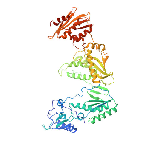
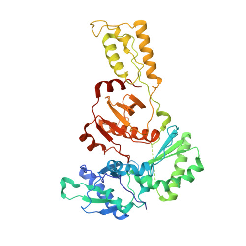
Complexes of HIV-1 RT, Nnrti and RNA/DNA Hybrid Reveal a Structure Compatible with RNA Degradation

Lapkouski, M.Tian, L.Miller, J.T.Le Grice, S.F.J.Yang, W.

(2013) Nat Struct Mol Biol 20: 230

  • DOI: https://doi.org/10.1038/nsmb.2485
  • Primary Citation Related Structures: 
    4B3O, 4B3P, 4B3Q

  • PubMed Abstract: 

    Hundreds of structures of type 1 human immunodeficiency virus (HIV-1) reverse transcriptase (RT) have been determined, but only one contains an RNA/DNA hybrid. Here we report three structures of HIV-1 RT complexed with a non-nucleotide RT inhibitor (NNRTI) and an RNA/DNA hybrid. In the presence of an NNRTI, the RNA/DNA structure differs from all prior nucleic acid-RT structures including the RNA/DNA hybrid. The enzyme structure also differs from all previous RT-DNA complexes. Thus, the hybrid has ready access to the RNase-H active site. These observations indicate that an RT-nucleic acid complex may adopt two structural states, one competent for DNA polymerization and the other for RNA degradation. RT mutations that confer drug resistance but are distant from the inhibitor-binding sites often map to the unique RT-hybrid interface that undergoes conformational changes between two catalytic states.


  • Organizational Affiliation
    • Laboratory of Molecular Biology, National Institute of Diabetes and Digestive and Kidney Diseases, National Institutes of Health, Bethesda, MD 20892, USA.

Macromolecule Content 

  • Total Structure Weight: 132.11 kDa 
  • Atom Count: 8,513 
  • Modeled Residue Count: 996 
  • Deposited Residue Count: 1,052 
  • Unique protein chains: 2
  • Unique nucleic acid chains: 2

Macromolecules


Find similar proteins by:|  3D Structure
Entity ID: 1
MoleculeChains  Sequence LengthOrganismDetailsImage
REVERSE TRANSCRIPTASE/RIBONUCLEASE H560Human immunodeficiency virus 1Mutation(s): 7 
EC: 2.7.7.49 (PDB Primary Data), 2.7.7.7 (PDB Primary Data), 3.1.26.13 (PDB Primary Data), 3.4.23.16 (PDB Primary Data), 3.1.13.2 (PDB Primary Data)
UniProt
Find proteins for P04585 (Human immunodeficiency virus type 1 group M subtype B (isolate HXB2))
Explore P04585 
Go to UniProtKB:  P04585
Entity Groups
Sequence Clusters30% Identity50% Identity70% Identity90% Identity95% Identity100% Identity
UniProt GroupP04585
Sequence Annotations
Expand
Reference Sequence
Find similar proteins by:|  3D Structure
Entity ID: 2
MoleculeChains  Sequence LengthOrganismDetailsImage
P51 RT441Human immunodeficiency virus 1Mutation(s): 3 
EC: 2.7.7.49 (PDB Primary Data), 2.7.7.7 (PDB Primary Data), 3.1.26.13 (PDB Primary Data), 3.4.23.16 (PDB Primary Data)
UniProt
Find proteins for P04585 (Human immunodeficiency virus type 1 group M subtype B (isolate HXB2))
Explore P04585 
Go to UniProtKB:  P04585
Entity Groups
Sequence Clusters30% Identity50% Identity70% Identity90% Identity95% Identity100% Identity
UniProt GroupP04585
Sequence Annotations
Expand
Reference Sequence
Find similar nucleic acids by:  Sequence
Entity ID: 3
MoleculeChains LengthOrganismImage
5'-D(*CP*GP*TP*AP*TP*GP*CP*CP*TP*AP*TP*AP*GP*TP *TP*AP*TP*TP*GP*TP*GP*GP*CP*C)-3'C [auth D]24synthetic construct
Sequence Annotations
Expand
Reference Sequence
Find similar nucleic acids by:  (by identity cutoff) 
Entity ID: 4
MoleculeChains LengthOrganismImage
5'-R(*AP*UP*GP*AP*3DRP*GP*GP*CP*CP*AP*CP*AP*AP*UP*AP *AP*CP*UP*AP*UP*AP*GP*GP*CP*AP*UP*A)-3'D [auth R]27synthetic construct
Sequence Annotations
Expand
Reference Sequence

Small Molecules

Binding Affinity Annotations 
IDSourceBinding Affinity
EFZ BindingDB:  4B3O Ki: min: 3, max: 3000 (nM) from 20 assay(s)
IC50: min: 0.09, max: 910 (nM) from 52 assay(s)
EC50: min: 1, max: 300 (nM) from 8 assay(s)

Experimental Data & Validation

Experimental Data

  • Method: X-RAY DIFFRACTION
  • Resolution: 3.30 Å
  • R-Value Free:  0.295 (Depositor), 0.298 (DCC) 
  • R-Value Work:  0.275 (Depositor), 0.277 (DCC) 
  • R-Value Observed: 0.276 (Depositor) 
Space Group: P 31 2 1
Unit Cell:
Length ( Å )Angle ( ˚ )
a = 164.598α = 90
b = 164.598β = 90
c = 129.03γ = 120
Software Package:
Software NamePurpose
PHENIXrefinement
XDSdata reduction
XSCALEdata scaling
MOLREPphasing

Structure Validation

View Full Validation Report


Ligand Structure Quality Assessment 


Entry History 

Deposition Data

Revision History  (Full details and data files)

  • Version 1.0: 2013-01-16
    Type: Initial release
  • Version 1.1: 2013-01-23
    Changes: Database references
  • Version 1.2: 2013-02-20
    Changes: Database references
  • Version 1.3: 2019-03-06
    Changes: Data collection, Derived calculations, Experimental preparation, Other
  • Version 1.4: 2019-05-15
    Changes: Data collection, Experimental preparation
  • Version 1.5: 2023-12-20
    Changes: Data collection, Database references, Derived calculations, Other, Refinement description