4AYC | pdb_00004ayc

RNF8 RING domain structure


Experimental Data Snapshot

  • Method: X-RAY DIFFRACTION
  • Resolution: 1.90 Å
  • R-Value Free: 
    0.229 (Depositor), 0.236 (DCC) 
  • R-Value Work: 
    0.200 (Depositor), 0.211 (DCC) 
  • R-Value Observed: 
    0.201 (Depositor) 

wwPDB Validation 3D Report Full Report

Validation slider image for 4AYC

Ligand Structure Quality Assessment 


This is version 1.3 of the entry. See complete history

Literature

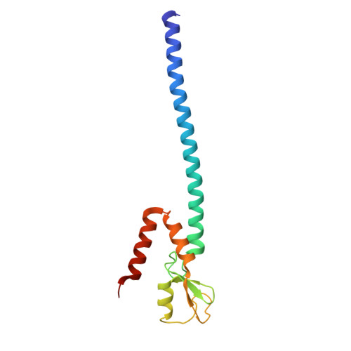
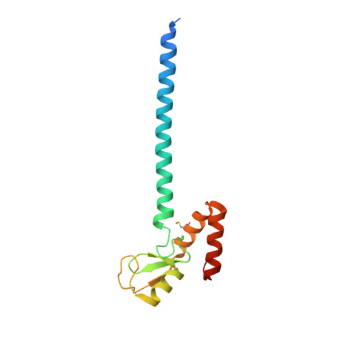
Rnf168 Ubiquitinates K13-15 on H2A/H2Ax to Drive DNA Damage Signaling

Mattiroli, F.Vissers, J.H.A.Van Dijk, W.J.Ikpa, P.Citterio, E.Vermeulen, W.Marteijn, J.A.Sixma, T.K.

(2012) Cell 150: 1182

  • DOI: https://doi.org/10.1016/j.cell.2012.08.005
  • Primary Citation Related Structures: 
    4AYC

  • PubMed Abstract: 

    Ubiquitin-dependent signaling during the DNA damage response (DDR) to double-strand breaks (DSBs) is initiated by two E3 ligases, RNF8 and RNF168, targeting histone H2A and H2AX. RNF8 is the first ligase recruited to the damage site, and RNF168 follows RNF8-dependent ubiquitination. This suggests that RNF8 initiates H2A/H2AX ubiquitination with K63-linked ubiquitin chains and RNF168 extends them. Here, we show that RNF8 is inactive toward nucleosomal H2A, whereas RNF168 catalyzes the monoubiquitination of the histones specifically on K13-15. Structure-based mutagenesis of RNF8 and RNF168 RING domains shows that a charged residue determines whether nucleosomal proteins are recognized. We find that K63 ubiquitin chains are conjugated to RNF168-dependent H2A/H2AX monoubiquitination at K13-15 and not on K118-119. Using a mutant of RNF168 unable to target histones but still catalyzing ubiquitin chains at DSBs, we show that ubiquitin chains per se are insufficient for signaling, but RNF168 target ubiquitination is required for DDR.


  • Organizational Affiliation
    • Division of Biochemistry and Center for Biomedical Genetics, Netherlands Cancer Institute, Plesmanlaan 121, 1066 CX Amsterdam, The Netherlands.

Macromolecule Content 

  • Total Structure Weight: 34.26 kDa 
  • Atom Count: 2,360 
  • Modeled Residue Count: 263 
  • Deposited Residue Count: 276 
  • Unique protein chains: 2

Macromolecules

Find similar proteins by:|  3D Structure
Entity ID: 1
MoleculeChains  Sequence LengthOrganismDetailsImage
E3 UBIQUITIN-PROTEIN LIGASE RNF8138Homo sapiensMutation(s): 0 
EC: 6.3.2 (PDB Primary Data), 2.3.2.27 (UniProt)
UniProt & NIH Common Fund Data Resources
Find proteins for O76064 (Homo sapiens)
Explore O76064 
Go to UniProtKB:  O76064
PHAROS:  O76064
GTEx:  ENSG00000112130 
Entity Groups
Sequence Clusters30% Identity50% Identity70% Identity90% Identity95% Identity100% Identity
UniProt GroupO76064
Sequence Annotations
Expand
Reference Sequence
Find similar proteins by:|  3D Structure
Entity ID: 2
MoleculeChains  Sequence LengthOrganismDetailsImage
E3 UBIQUITIN-PROTEIN LIGASE RNF8138Homo sapiensMutation(s): 0 
EC: 6.3.2 (PDB Primary Data), 2.3.2.27 (UniProt)
UniProt & NIH Common Fund Data Resources
Find proteins for O76064 (Homo sapiens)
Explore O76064 
Go to UniProtKB:  O76064
PHAROS:  O76064
GTEx:  ENSG00000112130 
Entity Groups
Sequence Clusters30% Identity50% Identity70% Identity90% Identity95% Identity100% Identity
UniProt GroupO76064
Sequence Annotations
Expand
Reference Sequence

Small Molecules

Ligands 5 Unique
IDChains Name / Formula / InChI Key2D Diagram3D Interactions
CPQ

Query on CPQ


Download:Ideal Coordinates CCD File
F [auth A]N,N-BIS(3-D-GLUCONAMIDOPROPYL)DEOXYCHOLAMIDE
C42 H75 N3 O15
OJSUWTDDXLCUFR-HGZMBBKESA-N
SO4

Query on SO4


Download:Ideal Coordinates CCD File
C [auth A],
D [auth A],
E [auth A]
SULFATE ION
O4 S
QAOWNCQODCNURD-UHFFFAOYSA-L
GOL

Query on GOL


Download:Ideal Coordinates CCD File
K [auth B]GLYCEROL
C3 H8 O3
PEDCQBHIVMGVHV-UHFFFAOYSA-N
ZN

Query on ZN


Download:Ideal Coordinates CCD File
G [auth A],
H [auth A],
I [auth B],
J [auth B]
ZINC ION
Zn
PTFCDOFLOPIGGS-UHFFFAOYSA-N
CL

Query on CL


Download:Ideal Coordinates CCD File
L [auth B]CHLORIDE ION
Cl
VEXZGXHMUGYJMC-UHFFFAOYSA-M
Modified Residues  1 Unique
IDChains TypeFormula2D DiagramParent
CSO
Query on CSO
B
L-PEPTIDE LINKINGC3 H7 N O3 SCYS

Experimental Data & Validation

Experimental Data

  • Method: X-RAY DIFFRACTION
  • Resolution: 1.90 Å
  • R-Value Free:  0.229 (Depositor), 0.236 (DCC) 
  • R-Value Work:  0.200 (Depositor), 0.211 (DCC) 
  • R-Value Observed: 0.201 (Depositor) 
Space Group: P 21 21 2
Unit Cell:
Length ( Å )Angle ( ˚ )
a = 47.374α = 90
b = 213.717β = 90
c = 34.102γ = 90
Software Package:
Software NamePurpose
REFMACrefinement
XDSdata reduction
SCALAdata scaling
SHELXDphasing

Structure Validation

View Full Validation Report


Ligand Structure Quality Assessment 


Entry History 

Deposition Data

Revision History  (Full details and data files)

  • Version 1.0: 2012-09-26
    Type: Initial release
  • Version 1.1: 2024-05-08
    Changes: Data collection, Database references, Derived calculations, Other
  • Version 1.2: 2024-09-11
    Changes: Derived calculations
  • Version 1.3: 2024-11-13
    Changes: Structure summary