4AUW

CRYSTAL STRUCTURE OF THE BZIP HOMODIMERIC MAFB IN COMPLEX WITH THE C- MARE BINDING SITE


Experimental Data Snapshot

  • Method: X-RAY DIFFRACTION
  • Resolution: 2.90 Å
  • R-Value Free: 0.290 
  • R-Value Work: 0.255 
  • R-Value Observed: 0.257 

wwPDB Validation   3D Report Full Report


Ligand Structure Quality Assessment 


This is version 1.1 of the entry. See complete history


Literature

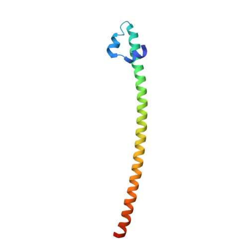
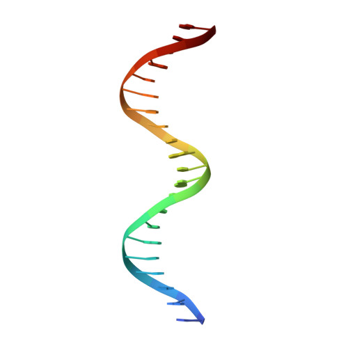
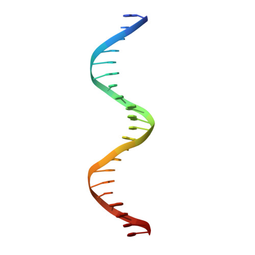
Expression, purification, crystallization and preliminary crystallographic analysis of the mouse transcription factor MafB in complex with its DNA-recognition motif Cmare

Textor, L.C.Wilmanns, M.Holton, S.J.

(2007) Acta Crystallogr Sect F Struct Biol Cryst Commun 63: 657-661

  • DOI: https://doi.org/10.1107/S1744309107031260
  • Primary Citation of Related Structures:  
    4AUW

  • PubMed Abstract: 

    The MafB transcription factor (residues 211-305) has been overexpressed in and purified from Escherichia coli. A protein-DNA complex between the MafB homodimer and the 21 bp Maf-recognition sequence known as Cmare has been successfully reconstituted in vitro and subsequently crystallized. The diffraction properties of the protein-DNA complex crystals were improved using a combination of protein-construct boundary optimization and targeted mutagenesis to promote crystal lattice stability. Both native and mercury-derivatized crystals have been prepared using these optimized conditions. The crystals belong to space group P4(1)2(1)2 or P4(3)2(1)2, with unit-cell parameters a = b = 94.8, c = 197.9 A. An anomalous difference Patterson map computed using data collected from crystals grown in the presence of HgCl(2) reveals four peaks. This corresponds to two copies of the protein-DNA complex in the asymmetric unit, with a solvent content of 62% and a Matthews coefficient of 3.22 A(3) Da(-1).


  • Organizational Affiliation

    EMBL DESY, Notkestrasse 85, D-22603 Hamburg, Germany.


Macromolecules

Find similar proteins by:  (by identity cutoff)  |  3D Structure
Entity ID: 1
MoleculeChains Sequence LengthOrganismDetailsImage
TRANSCRIPTION FACTOR MAFB
A, B, E, F
96Mus musculusMutation(s): 1 
UniProt
Find proteins for P54841 (Mus musculus)
Explore P54841 
Go to UniProtKB:  P54841
Entity Groups  
Sequence Clusters30% Identity50% Identity70% Identity90% Identity95% Identity100% Identity
UniProt GroupP54841
Sequence Annotations
Expand
  • Reference Sequence

Find similar nucleic acids by:  Sequence   |   3D Structure  

Entity ID: 2
MoleculeChains LengthOrganismImage
C-MARE BINDING SITE (5'-D(*AP*TP*AP*AP*TP*GP*CP*TP* GP*AP*CP*GP*TP*CP*AP*GP*CP*AP*AP*TP*T)-3')
C, H
21synthetic construct
Sequence Annotations
Expand
  • Reference Sequence

Find similar nucleic acids by:  Sequence   |   3D Structure  

Entity ID: 3
MoleculeChains LengthOrganismImage
C-MARE BINDING SITE (5'-D(*AP*TP*AP*AP*TP*GP*CP*TP* GP*AP*CP*GP*TP*CP*AP*GP*CP*AP*AP*TP*T)-3')
D, G
21synthetic construct
Sequence Annotations
Expand
  • Reference Sequence
Experimental Data & Validation

Experimental Data

  • Method: X-RAY DIFFRACTION
  • Resolution: 2.90 Å
  • R-Value Free: 0.290 
  • R-Value Work: 0.255 
  • R-Value Observed: 0.257 
  • Space Group: P 43 21 2
Unit Cell:
Length ( Å )Angle ( ˚ )
a = 94.964α = 90
b = 94.964β = 90
c = 200.111γ = 90
Software Package:
Software NamePurpose
REFMACrefinement

Structure Validation

View Full Validation Report


Ligand Structure Quality Assessment 


Entry History 

Deposition Data

Revision History  (Full details and data files)

  • Version 1.0: 2013-06-05
    Type: Initial release
  • Version 1.1: 2018-05-02
    Changes: Data collection, Database references