


The Structure of Bovine Chymosin in Complex with Pepstatin A
Langholm Jensen, J., Navarro Poulsen, J.C., Jacobsen, J., van den Brink, J.M., Qvist, K.B., Larsen, S.To be published.
Experimental Data Snapshot
Starting Model: experimental
View more details
wwPDB Validation 3D Report Full Report

Entity ID: 1 | |||||
|---|---|---|---|---|---|
| Molecule | Chains | Sequence Length | Organism | Details | Image |
| CHYMOSIN | 323 | Bos taurus | Mutation(s): 0 EC: 3.4.23.4 | 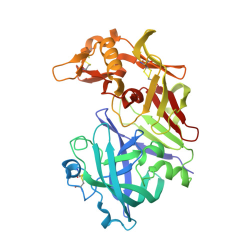 | |
UniProt | |||||
Entity Groups | |||||
| Sequence Clusters | 30% Identity50% Identity70% Identity90% Identity95% Identity100% Identity | ||||
| UniProt Group | P00794 | ||||
Sequence AnnotationsExpand | |||||
Reference Sequence | |||||
Entity ID: 2 | |||||
|---|---|---|---|---|---|
| Molecule | Chains | Sequence Length | Organism | Details | Image |
| PEPSTATIN A | 6 | Actinomyces | Mutation(s): 0 | 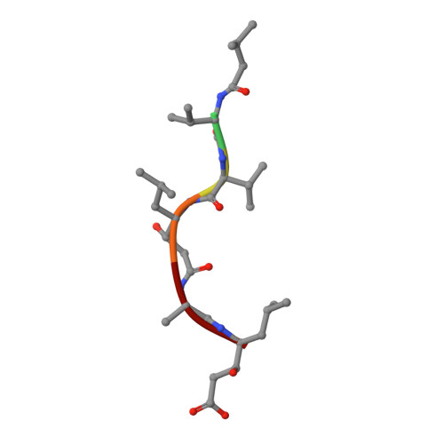 | |
| Ligands 3 Unique | |||||
|---|---|---|---|---|---|
| ID | Chains | Name / Formula / InChI Key | 2D Diagram | 3D Interactions | |
| CD Download:Ideal Coordinates CCD File | D [auth A] | CADMIUM ION Cd WLZRMCYVCSSEQC-UHFFFAOYSA-N |  | ||
| EDO Download:Ideal Coordinates CCD File | E [auth A], F [auth A], G [auth A], H [auth A] | 1,2-ETHANEDIOL C2 H6 O2 LYCAIKOWRPUZTN-UHFFFAOYSA-N |  | ||
| NA Download:Ideal Coordinates CCD File | C [auth A] | SODIUM ION Na FKNQFGJONOIPTF-UHFFFAOYSA-N |  | ||
| Length ( Å ) | Angle ( ˚ ) |
|---|---|
| a = 93.5 | α = 90 |
| b = 93.5 | β = 90 |
| c = 89 | γ = 120 |
| Software Name | Purpose |
|---|---|
| PHENIX | refinement |
| XDS | data reduction |
| XDS | data scaling |
| PHENIX | phasing |


