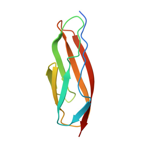
4AQO

CRYSTAL STRUCTURE OF THE CALCIUM BOUND PKD-like DOMAIN OF COLLAGENASE G FROM CLOSTRIDIUM HISTOLYTICUM AT 0.99 ANGSTROM RESOLUTION.


Experimental Data Snapshot

  • Method: X-RAY DIFFRACTION
  • Resolution: 0.99 Å
  • R-Value Free: 0.150 
  • R-Value Work: 0.124 
  • R-Value Observed: 0.125 

Starting Model: experimental
View more details

wwPDB Validation   3D Report Full Report


This is version 1.2 of the entry. See complete history


Literature

Structural Basis for Activity Regulation and Substrate Preference of Clostridial Collagenases G, H, and T.

Eckhard, U.Schonauer, E.Brandstetter, H.

(2013) J Biol Chem 288: 20184

  • DOI: https://doi.org/10.1074/jbc.M112.448548
  • Primary Citation of Related Structures:  
    4AQO, 4AR1, 4AR8, 4AR9, 4ARE, 4ARF

  • PubMed Abstract: 

    Clostridial collagenases are among the most efficient enzymes to degrade by far the most predominant protein in the biosphere. Here we present crystal structures of the peptidases of three clostridial collagenase isoforms (ColG, ColH, and ColT). The comparison of unliganded and liganded structures reveals a quaternary subdomain dynamics. In the unliganded ColH structure, this globular dynamics is modulated by an aspartate switch motion that binds to the catalytic zinc. We further identified a calcium binding site in proximity to the catalytic zinc. Both ions are required for full activity, explaining why calcium critically affects the enzymatic activity of clostridial collagenases. Our studies further reveal that loops close to the active site thus serve as characteristic substrate selectivity filter. These elements explain the distinct peptidolytic and collagenolytic activities of these enzymes and provide a rational framework to engineer collagenases with customized substrate specificity as well as for inhibitor design.


  • Organizational Affiliation

    Division of Structural Biology, Department of Molecular Biology, University of Salzburg, Billrothstrasse 11, A-5020 Salzburg, Austria.


Macromolecules
Find similar proteins by:  (by identity cutoff)  |  3D Structure
Entity ID: 1
MoleculeChains Sequence LengthOrganismDetailsImage
COLLAGENASE92Hathewaya histolyticaMutation(s): 0 
EC: 3.4.24.3
UniProt
Find proteins for Q9X721 (Hathewaya histolytica)
Explore Q9X721 
Go to UniProtKB:  Q9X721
Entity Groups  
Sequence Clusters30% Identity50% Identity70% Identity90% Identity95% Identity100% Identity
UniProt GroupQ9X721
Sequence Annotations
Expand
  • Reference Sequence
Small Molecules
Ligands 1 Unique
IDChains Name / Formula / InChI Key2D Diagram3D Interactions
CA
Query on CA

Download Ideal Coordinates CCD File 
B [auth A]CALCIUM ION
Ca
BHPQYMZQTOCNFJ-UHFFFAOYSA-N
Experimental Data & Validation

Experimental Data

  • Method: X-RAY DIFFRACTION
  • Resolution: 0.99 Å
  • R-Value Free: 0.150 
  • R-Value Work: 0.124 
  • R-Value Observed: 0.125 
  • Space Group: P 1 21 1
Unit Cell:
Length ( Å )Angle ( ˚ )
a = 19.753α = 90
b = 70.896β = 95.24
c = 23.379γ = 90
Software Package:
Software NamePurpose
REFMACrefinement
MOSFLMdata reduction
SCALAdata scaling
PHASERphasing

Structure Validation

View Full Validation Report


Entry History 

Deposition Data

Revision History  (Full details and data files)

  • Version 1.0: 2013-06-05
    Type: Initial release
  • Version 1.1: 2013-07-31
    Changes: Database references
  • Version 1.2: 2023-12-20
    Changes: Data collection, Database references, Derived calculations, Other, Refinement description