4AQ6

substrate bound homogentisate 1,2-dioxygenase


Experimental Data Snapshot

  • Method: X-RAY DIFFRACTION
  • Resolution: 1.98 Å
  • R-Value Free: 0.216 
  • R-Value Work: 0.161 
  • R-Value Observed: 0.164 

wwPDB Validation   3D Report Full Report


Ligand Structure Quality Assessment 


This is version 1.4 of the entry. See complete history


Literature

Visualizing the Substrate-, Superoxo-, Alkylperoxo- and Product-Bound States at the Non-Heme Fe(II) Site of Homogentisate Dioxygenase

Jeoung, J.-H.Bommer, M.Lin, T.-Y.Dobbek, H.

(2013) Proc Natl Acad Sci U S A 110: 12625

  • DOI: https://doi.org/10.1073/pnas.1302144110
  • Primary Citation of Related Structures:  
    3ZDS, 4AQ2, 4AQ6

  • PubMed Abstract: 

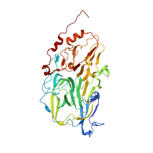
    Homogentisate 1,2-dioxygenase (HGDO) uses a mononuclear nonheme Fe(2+) to catalyze the oxidative ring cleavage in the degradation of Tyr and Phe by producing maleylacetoacetate from homogentisate (2,5-dihydroxyphenylacetate). Here, we report three crystal structures of HGDO, revealing five different steps in its reaction cycle at 1.7-1.98 Å resolution. The resting state structure displays an octahedral coordination for Fe(2+) with two histidine residues (His331 and His367), a bidentate carboxylate ligand (Glu337), and two water molecules. Homogentisate binds as a monodentate ligand to Fe(2+), and its interaction with Tyr346 invokes the folding of a loop over the active site, effectively shielding it from solvent. Binding of homogentisate is driven by enthalpy and is entropically disfavored as shown by anoxic isothermal titration calorimetry. Three different reaction cycle intermediates have been trapped in different HGDO subunits of a single crystal showing the influence of crystal packing interactions on the course of enzymatic reactions. The observed superoxo:semiquinone-, alkylperoxo-, and product-bound intermediates have been resolved in a crystal grown anoxically with homogentisate, which was subsequently incubated with dioxygen. We demonstrate that, despite different folds, active site architectures, and Fe(2+) coordination, extradiol dioxygenases can proceed through the same principal reaction intermediates to catalyze the O2-dependent cleavage of aromatic rings. Thus, convergent evolution of nonhomologous enzymes using the 2-His-1-carboxylate facial triad motif developed different solutions to stabilize closely related intermediates in unlike environments.


  • Organizational Affiliation

    Institut für Biologie, Strukturbiologie/Biochemie, Humboldt-Universität zu Berlin, 10099 Berlin, Germany.


Macromolecules
Find similar proteins by:  (by identity cutoff)  |  3D Structure
Entity ID: 1
MoleculeChains Sequence LengthOrganismDetailsImage
HOMOGENTISATE 1,2-DIOXYGENASE
A, B, C, D, E
A, B, C, D, E, F, G, H, I, J, K, L
433Pseudomonas putida KT2440Mutation(s): 0 
EC: 1.13.11.5
UniProt
Find proteins for Q88E47 (Pseudomonas putida (strain ATCC 47054 / DSM 6125 / CFBP 8728 / NCIMB 11950 / KT2440))
Explore Q88E47 
Go to UniProtKB:  Q88E47
Entity Groups  
Sequence Clusters30% Identity50% Identity70% Identity90% Identity95% Identity100% Identity
UniProt GroupQ88E47
Sequence Annotations
Expand
  • Reference Sequence
Small Molecules
Ligands 3 Unique
IDChains Name / Formula / InChI Key2D Diagram3D Interactions
B3P
Query on B3P

Download Ideal Coordinates CCD File 
IA [auth K]2-[3-(2-HYDROXY-1,1-DIHYDROXYMETHYL-ETHYLAMINO)-PROPYLAMINO]-2-HYDROXYMETHYL-PROPANE-1,3-DIOL
C11 H26 N2 O6
HHKZCCWKTZRCCL-UHFFFAOYSA-N
OMD
Query on OMD

Download Ideal Coordinates CCD File 
BA [auth H]
DA [auth I]
FA [auth J]
HA [auth K]
KA [auth L]
BA [auth H],
DA [auth I],
FA [auth J],
HA [auth K],
KA [auth L],
N [auth A],
P [auth B],
R [auth C],
T [auth D],
V [auth E],
X [auth F],
Z [auth G]
2-(3,6-DIHYDROXYPHENYL)ACETIC ACID
C8 H8 O4
IGMNYECMUMZDDF-UHFFFAOYSA-N
FE
Query on FE

Download Ideal Coordinates CCD File 
AA [auth H]
CA [auth I]
EA [auth J]
GA [auth K]
JA [auth L]
AA [auth H],
CA [auth I],
EA [auth J],
GA [auth K],
JA [auth L],
M [auth A],
O [auth B],
Q [auth C],
S [auth D],
U [auth E],
W [auth F],
Y [auth G]
FE (III) ION
Fe
VTLYFUHAOXGGBS-UHFFFAOYSA-N
Binding Affinity Annotations 
IDSourceBinding Affinity
OMD PDBBind:  4AQ6 Kd: 1.58e+4 (nM) from 1 assay(s)
Experimental Data & Validation

Experimental Data

  • Method: X-RAY DIFFRACTION
  • Resolution: 1.98 Å
  • R-Value Free: 0.216 
  • R-Value Work: 0.161 
  • R-Value Observed: 0.164 
  • Space Group: P 1
Unit Cell:
Length ( Å )Angle ( ˚ )
a = 93.393α = 87.69
b = 93.714β = 80.42
c = 162.984γ = 68.39
Software Package:
Software NamePurpose
PHENIXrefinement
XDSdata reduction
XSCALEdata scaling
PHASERphasing

Structure Validation

View Full Validation Report


Ligand Structure Quality Assessment 


Entry History 

Deposition Data

Revision History  (Full details and data files)

  • Version 1.0: 2013-04-24
    Type: Initial release
  • Version 1.1: 2013-07-24
    Changes: Database references, Other
  • Version 1.2: 2013-08-14
    Changes: Database references
  • Version 1.3: 2019-03-06
    Changes: Data collection, Experimental preparation, Other
  • Version 1.4: 2023-12-20
    Changes: Data collection, Database references, Derived calculations, Other, Refinement description