4ANP

Crystal structure of human phenylalanine hydroxylase in complex with a pharmacological chaperone


Experimental Data Snapshot

  • Method: X-RAY DIFFRACTION
  • Resolution: 2.11 Å
  • R-Value Free: 0.237 
  • R-Value Work: 0.204 
  • R-Value Observed: 0.205 

Starting Model: experimental
View more details

wwPDB Validation   3D Report Full Report


Ligand Structure Quality Assessment 


This is version 1.2 of the entry. See complete history


Literature

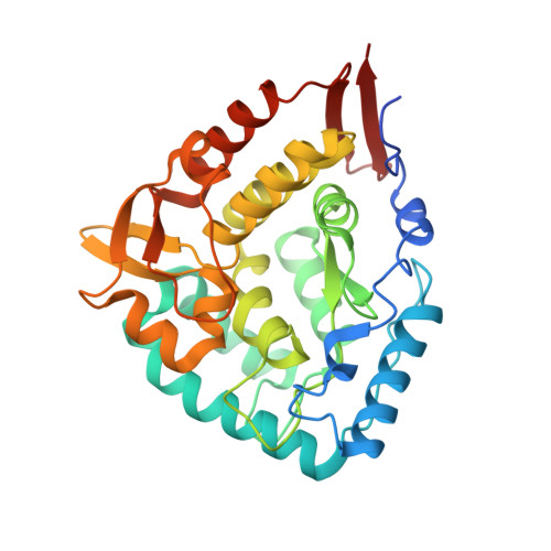
Structural and Mechanistic Basis of the Interaction between a Pharmacological Chaperone and Human Phenylalanine Hydroxylase.

Torreblanca, R.Lira-Navarrete, E.Sancho, J.Hurtado-Guerrero, R.

(2012) Chembiochem 13: 1266

  • DOI: https://doi.org/10.1002/cbic.201200188
  • Primary Citation of Related Structures:  
    4ANP

  • PubMed Abstract: 

    Not without a chaperone: Pharmacological chaperones are designed to bind and ideally stabilise their target protein. Here, we elucidate the molecular mechanism of a potential pharmacological chaperone to treat phenylketonuria. The crystal structure of human phenylalanine hydroxylase with compound IV may help in the rational design of more efficient compounds to treat this disease.


  • Organizational Affiliation

    Department of Biochemistry and Molecular and Cellular Biology, Institute of Biocomputation and Physics of Complex Systems (BIFI), University of Zaragoza, BIFI-IQFR (CSIC) Joint Unit, Pedro Cerbuna 12, Mariano Esquillor s/n, Campus Rio Ebro, Edificio I+D, Spain.


Macromolecules
Find similar proteins by:  (by identity cutoff)  |  3D Structure
Entity ID: 1
MoleculeChains Sequence LengthOrganismDetailsImage
PHENYLALANINE-4-HYDROXYLASE324Homo sapiensMutation(s): 0 
EC: 1.14.16.1
UniProt & NIH Common Fund Data Resources
Find proteins for P00439 (Homo sapiens)
Explore P00439 
Go to UniProtKB:  P00439
GTEx:  ENSG00000171759 
Entity Groups  
Sequence Clusters30% Identity50% Identity70% Identity90% Identity95% Identity100% Identity
UniProt GroupP00439
Sequence Annotations
Expand
  • Reference Sequence
Small Molecules
Ligands 2 Unique
IDChains Name / Formula / InChI Key2D Diagram3D Interactions
3QI
Query on 3QI

Download Ideal Coordinates CCD File 
C [auth A]5,6-DIMETHYL-3-(4-METHYL-2-PYRIDINYL)-2-THIOXO-2,3-DIHYDROTHIENO[2,3- D]PYRIMIDIN-4(1H)-ONE
C14 H13 N3 O S2
NYDQMBWQPMNQIZ-UHFFFAOYSA-N
FE
Query on FE

Download Ideal Coordinates CCD File 
B [auth A]FE (III) ION
Fe
VTLYFUHAOXGGBS-UHFFFAOYSA-N
Experimental Data & Validation

Experimental Data

  • Method: X-RAY DIFFRACTION
  • Resolution: 2.11 Å
  • R-Value Free: 0.237 
  • R-Value Work: 0.204 
  • R-Value Observed: 0.205 
  • Space Group: C 2 2 21
Unit Cell:
Length ( Å )Angle ( ˚ )
a = 66.283α = 90
b = 107.511β = 90
c = 124.55γ = 90
Software Package:
Software NamePurpose
REFMACrefinement
SAINTdata reduction
SADABSdata scaling
MOLREPphasing

Structure Validation

View Full Validation Report


Ligand Structure Quality Assessment 


Entry History 

Deposition Data

Revision History  (Full details and data files)

  • Version 1.0: 2012-04-11
    Type: Initial release
  • Version 1.1: 2012-07-25
    Changes: Database references
  • Version 1.2: 2023-12-20
    Changes: Data collection, Database references, Derived calculations, Other, Refinement description