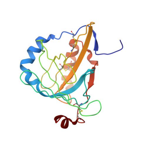
4ALO

STRUCTURE AND PROPERTIES OF H1 CRUSTACYANIN FROM LOBSTER HOMARUS AMERICANUS


Experimental Data Snapshot

  • Method: X-RAY DIFFRACTION
  • Resolution: 2.37 Å
  • R-Value Free: 0.266 
  • R-Value Work: 0.222 
  • R-Value Observed: 0.225 

Starting Model: experimental
View more details

wwPDB Validation   3D Report Full Report


This is version 1.2 of the entry. See complete history


Literature

Structural Characterization of Recombinant Crustacyanin Subunits from the Lobster Homarus Americanus.

Ferrari, M.Folli, C.Pincolini, E.Mcclintock, T.S.Rossle, M.Berni, R.Cianci, M.

(2012) Acta Crystallogr Sect F Struct Biol Cryst Commun 68: 846

  • DOI: https://doi.org/10.1107/S1744309112026103
  • Primary Citation of Related Structures:  
    4ALO

  • PubMed Abstract: 

    Crustacean crustacyanin proteins are linked to the production and modification of carapace colour, with direct implications for fitness and survival. Here, the structural and functional properties of the two recombinant crustacyanin subunits H(1) and H(2) from the American lobster Homarus americanus are reported. The two subunits are structurally highly similar to the corresponding natural apo crustacyanin CRTC and CRTA subunits from the European lobster H. gammarus. Reconstitution studies of the recombinant crustacyanin proteins H(1) and H(2) with astaxanthin reproduced the bathochromic shift of 85-95 nm typical of the natural crustacyanin subunits from H. gammarus in complex with astaxanthin. Moreover, correlations between the presence of crustacyanin genes in crustacean species and the resulting carapace colours with the spectral properties of the subunits in complex with astaxanthin confirmed this genotype-phenotype linkage.


  • Organizational Affiliation

    Department of Biochemistry and Molecular Biology, University of Parma, Parco Area delle Scienze 23/A, 43124 Parma, Italy.


Macromolecules
Find similar proteins by:  (by identity cutoff)  |  3D Structure
Entity ID: 1
MoleculeChains Sequence LengthOrganismDetailsImage
H1 APOCRUSTACYANIN
A, B
181Homarus americanusMutation(s): 0 
UniProt
Find proteins for J3QW35 (Homarus americanus)
Explore J3QW35 
Go to UniProtKB:  J3QW35
Entity Groups  
Sequence Clusters30% Identity50% Identity70% Identity90% Identity95% Identity100% Identity
UniProt GroupJ3QW35
Sequence Annotations
Expand
  • Reference Sequence
Small Molecules
Ligands 3 Unique
IDChains Name / Formula / InChI Key2D Diagram3D Interactions
MPD
Query on MPD

Download Ideal Coordinates CCD File 
C [auth A],
D [auth A],
H [auth A],
I [auth B],
J [auth B]
(4S)-2-METHYL-2,4-PENTANEDIOL
C6 H14 O2
SVTBMSDMJJWYQN-YFKPBYRVSA-N
SO4
Query on SO4

Download Ideal Coordinates CCD File 
E [auth A],
K [auth B],
M [auth B],
N [auth B],
O [auth B]
SULFATE ION
O4 S
QAOWNCQODCNURD-UHFFFAOYSA-L
NA
Query on NA

Download Ideal Coordinates CCD File 
F [auth A],
G [auth A],
L [auth B]
SODIUM ION
Na
FKNQFGJONOIPTF-UHFFFAOYSA-N
Experimental Data & Validation

Experimental Data

  • Method: X-RAY DIFFRACTION
  • Resolution: 2.37 Å
  • R-Value Free: 0.266 
  • R-Value Work: 0.222 
  • R-Value Observed: 0.225 
  • Space Group: P 21 21 21
Unit Cell:
Length ( Å )Angle ( ˚ )
a = 40.368α = 90
b = 78.423β = 90
c = 105.625γ = 90
Software Package:
Software NamePurpose
REFMACrefinement
DENZOdata reduction
SCALEPACKdata scaling
MOLREPphasing

Structure Validation

View Full Validation Report


Entry History 

Deposition Data

Revision History  (Full details and data files)

  • Version 1.0: 2012-08-08
    Type: Initial release
  • Version 1.1: 2012-08-15
    Changes: Database references
  • Version 1.2: 2023-12-20
    Changes: Data collection, Database references, Derived calculations, Other, Refinement description