4AJ5

Crystal structure of the Ska core complex


Experimental Data Snapshot

  • Method: X-RAY DIFFRACTION
  • Resolution: 3.32 Å
  • R-Value Free: 0.280 
  • R-Value Work: 0.234 
  • R-Value Observed: 0.237 

wwPDB Validation   3D Report Full Report


This is version 1.0 of the entry. See complete history


Literature

Structural and Functional Organization of the Ska Complex, a Key Component of the Kinetochore-Microtubule Interface.

Jeyaprakash, A.A.Santamaria, A.Jayachandran, U.Chan, Y.W.Benda, C.Nigg, E.A.Conti, E.

(2012) Mol Cell 46: 274

  • DOI: https://doi.org/10.1016/j.molcel.2012.03.005
  • Primary Citation of Related Structures:  
    4AJ5

  • PubMed Abstract: 

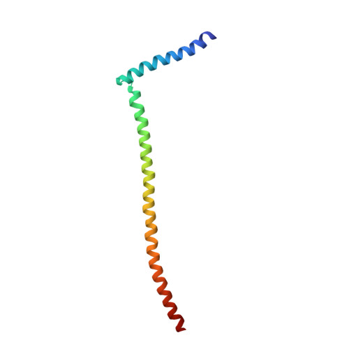
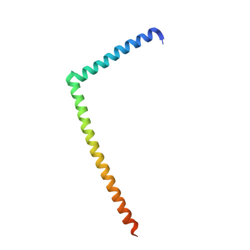
    The Ska complex is an essential mitotic component required for accurate cell division in human cells. It is composed of three subunits that function together to establish stable kinetochore-microtubule interactions in concert with the Ndc80 network. We show that the structure of the Ska core complex is a W-shaped dimer of coiled coils, formed by intertwined interactions between Ska1, Ska2, and Ska3. The C-terminal domains of Ska1 and Ska3 protrude at each end of the homodimer, bind microtubules in vitro when connected to the central core, and are essential in vivo. Mutations disrupting the central coiled coil or the dimerization interface result in chromosome congression failure followed by cell death. The Ska complex is thus endowed with bipartite and cooperative tubulin-binding properties at the ends of a 350 Å-long molecule. We discuss how this symmetric architecture might complement and stabilize the Ndc80-microtubule attachments with analogies to the yeast Dam1/DASH complex.


  • Organizational Affiliation

    Max-Planck-Institute of Biochemistry, Department of Structural Cell Biology, Martinsried, Germany.


Macromolecules
Find similar proteins by:  (by identity cutoff)  |  3D Structure
Entity ID: 1
MoleculeChains Sequence LengthOrganismDetailsImage
SPINDLE AND KINETOCHORE-ASSOCIATED PROTEIN 3101Homo sapiensMutation(s): 1 
UniProt & NIH Common Fund Data Resources
Find proteins for Q8IX90 (Homo sapiens)
Explore Q8IX90 
Go to UniProtKB:  Q8IX90
PHAROS:  Q8IX90
GTEx:  ENSG00000165480 
Entity Groups  
Sequence Clusters30% Identity50% Identity70% Identity90% Identity95% Identity100% Identity
UniProt GroupQ8IX90
Sequence Annotations
Expand
  • Reference Sequence
Find similar proteins by:  (by identity cutoff)  |  3D Structure
Entity ID: 2
MoleculeChains Sequence LengthOrganismDetailsImage
SPINDLE AND KINETOCHORE-ASSOCIATED PROTEIN 191Homo sapiensMutation(s): 0 
UniProt & NIH Common Fund Data Resources
Find proteins for Q96BD8 (Homo sapiens)
Explore Q96BD8 
Go to UniProtKB:  Q96BD8
PHAROS:  Q96BD8
GTEx:  ENSG00000154839 
Entity Groups  
Sequence Clusters30% Identity50% Identity70% Identity90% Identity95% Identity100% Identity
UniProt GroupQ96BD8
Sequence Annotations
Expand
  • Reference Sequence
Find similar proteins by:  (by identity cutoff)  |  3D Structure
Entity ID: 3
MoleculeChains Sequence LengthOrganismDetailsImage
SPINDLE AND KINETOCHORE-ASSOCIATED PROTEIN 2123Homo sapiensMutation(s): 0 
UniProt & NIH Common Fund Data Resources
Find proteins for Q8WVK7 (Homo sapiens)
Explore Q8WVK7 
Go to UniProtKB:  Q8WVK7
PHAROS:  Q8WVK7
GTEx:  ENSG00000182628 
Entity Groups  
Sequence Clusters30% Identity50% Identity70% Identity90% Identity95% Identity100% Identity
UniProt GroupQ8WVK7
Sequence Annotations
Expand
  • Reference Sequence
Experimental Data & Validation

Experimental Data

  • Method: X-RAY DIFFRACTION
  • Resolution: 3.32 Å
  • R-Value Free: 0.280 
  • R-Value Work: 0.234 
  • R-Value Observed: 0.237 
  • Space Group: C 1 2 1
Unit Cell:
Length ( Å )Angle ( ˚ )
a = 174.4α = 90
b = 193.121β = 95.21
c = 198.267γ = 90
Software Package:
Software NamePurpose
PHENIXrefinement
XDSdata reduction
SCALAdata scaling
PHENIXphasing

Structure Validation

View Full Validation Report


Entry History 

Deposition Data

Revision History  (Full details and data files)

  • Version 1.0: 2012-05-23
    Type: Initial release