4AG9 | pdb_00004ag9

C. elegans glucosamine-6-phosphate N-acetyltransferase (GNA1): ternary complex with coenzyme A and GlcNAc


Experimental Data Snapshot

  • Method: X-RAY DIFFRACTION
  • Resolution: 1.76 Å
  • R-Value Free: 
    0.232 (Depositor), 0.212 (DCC) 
  • R-Value Work: 
    0.190 (Depositor), 0.189 (DCC) 
  • R-Value Observed: 
    0.191 (Depositor) 

Starting Model: experimental
View more details

wwPDB Validation 3D Report Full Report

Validation slider image for 4AG9

Ligand Structure Quality Assessment 


This is version 1.3 of the entry. See complete history

Literature

Structural and Biochemical Characterization of a Trapped Coenzyme a Adduct of Caenorhabditis Elegans Glucosamine-6-Phosphate N-Acetyltransferase 1.

Dorfmueller, H.C.Fang, W.Rao, F.V.Blair, D.E.Attrill, H.Van Aalten, D.M.F.

(2012) Acta Crystallogr D Biol Crystallogr 68: 1019

  • DOI: https://doi.org/10.1107/S0907444912019592
  • Primary Citation Related Structures: 
    4AG7, 4AG9

  • PubMed Abstract: 

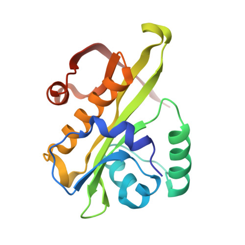
    Glucosamine-6-phosphate N-acetyltransferase 1 (GNA1) produces GlcNAc-6-phosphate from GlcN-6-phosphate and acetyl coenzyme A. Early mercury-labelling experiments implicated a conserved cysteine in the reaction mechanism, whereas recent structural data appear to support a mechanism in which this cysteine plays no role. Here, two crystal structures of Caenorhabditis elegans GNA1 are reported, revealing an unusual covalent complex between this cysteine and the coenzyme A product. Mass-spectrometric and reduction studies showed that this inactive covalent complex can be reactivated through reduction, yet mutagenesis of the cysteine supports a previously reported bi-bi mechanism. The data unify the apparently contradictory earlier reports on the role of a cysteine in the GNA1 active site.


  • Organizational Affiliation
    • Division of Molecular Microbiology, College of Life Sciences, University of Dundee, Dundee DD1 5EH, Scotland.

Macromolecule Content 

  • Total Structure Weight: 39.22 kDa 
  • Atom Count: 3,133 
  • Modeled Residue Count: 328 
  • Deposited Residue Count: 330 
  • Unique protein chains: 1

Macromolecules

Find similar proteins by:|  3D Structure
Entity ID: 1
MoleculeChains  Sequence LengthOrganismDetailsImage
GLUCOSAMINE-6-PHOSPHATE N-ACETYLTRANSFERASE
A, B
165Caenorhabditis elegansMutation(s): 0 
EC: 2.3.1.4
UniProt
Find proteins for Q17427 (Caenorhabditis elegans)
Explore Q17427 
Go to UniProtKB:  Q17427
Entity Groups
Sequence Clusters30% Identity50% Identity70% Identity90% Identity95% Identity100% Identity
UniProt GroupQ17427
Sequence Annotations
Expand
Reference Sequence

Experimental Data & Validation

Experimental Data

  • Method: X-RAY DIFFRACTION
  • Resolution: 1.76 Å
  • R-Value Free:  0.232 (Depositor), 0.212 (DCC) 
  • R-Value Work:  0.190 (Depositor), 0.189 (DCC) 
  • R-Value Observed: 0.191 (Depositor) 
Space Group: P 21 21 21
Unit Cell:
Length ( Å )Angle ( ˚ )
a = 48.872α = 90
b = 53.353β = 90
c = 135.096γ = 90
Software Package:
Software NamePurpose
REFMACrefinement
DENZOdata reduction
SCALEPACKdata scaling
MOLREPphasing

Structure Validation

View Full Validation Report


Ligand Structure Quality Assessment 


Entry History 

Deposition Data

Revision History  (Full details and data files)

  • Version 1.0: 2012-07-25
    Type: Initial release
  • Version 1.1: 2012-08-15
    Changes: Database references
  • Version 1.2: 2020-07-29
    Type: Remediation
    Reason: Carbohydrate remediation
    Changes: Data collection, Derived calculations, Other, Structure summary
  • Version 1.3: 2023-12-20
    Changes: Data collection, Database references, Refinement description, Structure summary