


Structural Determinants of Aromatic Ring Reduction in the Xenobiotic Reductase B from Pseudomonas Putida
Carvalho, A.L., Mukhopadhyay, A., Bonifacio, C., Romao, M.J., Bursakov, S., Kladova, A., Ramos, J.L., Van Dillewijn, P.To be published.
Experimental Data Snapshot
Entity ID: 1 | |||||
|---|---|---|---|---|---|
| Molecule | Chains | Sequence Length | Organism | Details | Image |
| XENOBIOTIC REDUCTASE B | 362 | Pseudomonas putida KT2440 | Mutation(s): 0 | 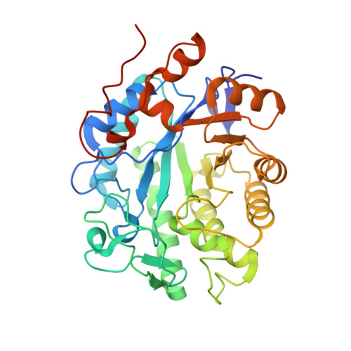 | |
UniProt | |||||
Entity Groups | |||||
| Sequence Clusters | 30% Identity50% Identity70% Identity90% Identity95% Identity100% Identity | ||||
| UniProt Group | Q88PD0 | ||||
Sequence AnnotationsExpand | |||||
Reference Sequence | |||||
| Ligands 4 Unique | |||||
|---|---|---|---|---|---|
| ID | Chains | Name / Formula / InChI Key | 2D Diagram | 3D Interactions | |
| FMN Download:Ideal Coordinates CCD File | C [auth A], H [auth B] | FLAVIN MONONUCLEOTIDE C17 H21 N4 O9 P FVTCRASFADXXNN-SCRDCRAPSA-N |  | ||
| TNL Download:Ideal Coordinates CCD File | G [auth A], J [auth B] | 2,4,6-TRINITROTOLUENE C7 H5 N3 O6 SPSSULHKWOKEEL-UHFFFAOYSA-N |  | ||
| SO4 Download:Ideal Coordinates CCD File | E [auth A], F [auth A], I [auth B] | SULFATE ION O4 S QAOWNCQODCNURD-UHFFFAOYSA-L |  | ||
| GOL Download:Ideal Coordinates CCD File | D [auth A] | GLYCEROL C3 H8 O3 PEDCQBHIVMGVHV-UHFFFAOYSA-N |  | ||
| Length ( Å ) | Angle ( ˚ ) |
|---|---|
| a = 67.18 | α = 90 |
| b = 105.32 | β = 90 |
| c = 106.8 | γ = 90 |
| Software Name | Purpose |
|---|---|
| PHENIX | refinement |
| MOSFLM | data reduction |
| SCALA | data scaling |
| PHASER | phasing |


