4A5Y

Intermediate state of human kinesin Eg5 in complex with Ispinesib


Experimental Data Snapshot

  • Method: X-RAY DIFFRACTION
  • Resolution: 2.45 Å
  • R-Value Free: 0.268 
  • R-Value Work: 0.201 
  • R-Value Observed: 0.204 

wwPDB Validation   3D Report Full Report


Ligand Structure Quality Assessment 


This is version 1.3 of the entry. See complete history


Literature

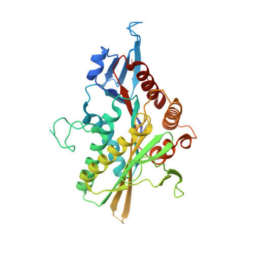
"Snapshots" of Ispinesib-Induced Conformational Changes in the Mitotic Kinesin Eg5.

Kaan, H.Y.K.Major, J.Tkocz, K.Kozielski, F.Rosenfeld, S.S.

(2013) J Biol Chem 288: 18588

  • DOI: https://doi.org/10.1074/jbc.M113.462648
  • Primary Citation of Related Structures:  
    4A5Y

  • PubMed Abstract: 

    Kinesins comprise a superfamily of molecular motors that drive a wide variety of cellular physiologies, from cytoplasmic transport to formation of the bipolar spindle in mitosis. These differing roles are reflected in corresponding polymorphisms in key kinesin structural elements. One of these is a unique loop and stem motif found in all kinesins and referred to as loop 5 (L5). This loop is longest in the mitotic kinesin Eg5 and is the target for a number of small molecule inhibitors, including ispinesib, which is being used in clinical trials in patients with cancer. In this study, we have used x-ray crystallography to identify a new structure of an Eg5-ispinesib complex and have combined this with transient state kinetics to identify a plausible sequence of conformational changes that occur in response to ispinesib binding. Our results demonstrate that ispinesib-induced structural changes in L5 from Eg5 lead to subsequent changes in the conformation of the switch II loop and helix and in the neck linker. We conclude that L5 in Eg5 simultaneously regulates the structure of both the ATP binding site and the motor's mechanical elements that generate force.


  • Organizational Affiliation

    Beatson Institute for Cancer Research, Garscube Estate, Switchback Road, Bearsden, Glasgow G61 1BD, Scotland, United Kingdom.


Macromolecules
Find similar proteins by:  (by identity cutoff)  |  3D Structure
Entity ID: 1
MoleculeChains Sequence LengthOrganismDetailsImage
KINESIN-LIKE PROTEIN KIF11
A, B, C
368Homo sapiensMutation(s): 0 
UniProt & NIH Common Fund Data Resources
Find proteins for P52732 (Homo sapiens)
Explore P52732 
Go to UniProtKB:  P52732
PHAROS:  P52732
GTEx:  ENSG00000138160 
Entity Groups  
Sequence Clusters30% Identity50% Identity70% Identity90% Identity95% Identity100% Identity
UniProt GroupP52732
Sequence Annotations
Expand
  • Reference Sequence
Small Molecules
Ligands 5 Unique
IDChains Name / Formula / InChI Key2D Diagram3D Interactions
G7X
Query on G7X

Download Ideal Coordinates CCD File 
F [auth A],
H [auth B],
N [auth C]
ISPINESIB MESILATE
C30 H33 Cl N4 O2
QJZRFPJCWMNVAV-HHHXNRCGSA-N
ADP
Query on ADP

Download Ideal Coordinates CCD File 
D [auth A],
G [auth B],
I [auth C]
ADENOSINE-5'-DIPHOSPHATE
C10 H15 N5 O10 P2
XTWYTFMLZFPYCI-KQYNXXCUSA-N
PO4
Query on PO4

Download Ideal Coordinates CCD File 
K [auth C],
L [auth C]
PHOSPHATE ION
O4 P
NBIIXXVUZAFLBC-UHFFFAOYSA-K
CL
Query on CL

Download Ideal Coordinates CCD File 
E [auth A],
M [auth C]
CHLORIDE ION
Cl
VEXZGXHMUGYJMC-UHFFFAOYSA-M
MG
Query on MG

Download Ideal Coordinates CCD File 
J [auth C]MAGNESIUM ION
Mg
JLVVSXFLKOJNIY-UHFFFAOYSA-N
Binding Affinity Annotations 
IDSourceBinding Affinity
G7X BindingDB:  4A5Y Ki: min: 2.3, max: 2.5 (nM) from 2 assay(s)
IC50: min: 1.2, max: 47 (nM) from 3 assay(s)
Experimental Data & Validation

Experimental Data

  • Method: X-RAY DIFFRACTION
  • Resolution: 2.45 Å
  • R-Value Free: 0.268 
  • R-Value Work: 0.201 
  • R-Value Observed: 0.204 
  • Space Group: P 21 21 21
Unit Cell:
Length ( Å )Angle ( ˚ )
a = 85.63α = 90
b = 91.93β = 90
c = 163.9γ = 90
Software Package:
Software NamePurpose
PHENIXrefinement
iMOSFLMdata reduction
SCALAdata scaling
MOLREPphasing

Structure Validation

View Full Validation Report


Ligand Structure Quality Assessment 


Entry History 

Deposition Data

Revision History  (Full details and data files)

  • Version 1.0: 2012-11-07
    Type: Initial release
  • Version 1.1: 2013-05-15
    Changes: Other, Structure summary
  • Version 1.2: 2015-05-20
    Changes: Database references
  • Version 1.3: 2023-12-20
    Changes: Data collection, Database references, Derived calculations, Other, Refinement description