Dissection of the Network of Interactions that Links RNA Processing with Glycolysis in the Bacillus Subtilis Degradosome.
Newman, J.A., Hewitt, L., Rodrigues, C., Solovyova, A.S., Harwood, C.R., Lewis, R.J.(2012) J Mol Biol 416: 121
- PubMed: 22198292 Search on PubMed
- DOI: https://doi.org/10.1016/j.jmb.2011.12.024
- Primary Citation Related Structures:
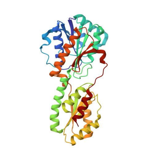
4A3R, 4A3S - PubMed Abstract:
The RNA degradosome is a multiprotein macromolecular complex that is involved in the degradation of messenger RNA in bacteria. The composition of this complex has been found to display a high degree of evolutionary divergence, which may reflect the adaptation of species to different environments. Recently, a degradosome-like complex identified in Bacillus subtilis was found to be distinct from those found in proteobacteria, the degradosomes of which are assembled around the unstructured C-terminus of ribonuclease E, a protein not present in B. subtilis. In this report, we have investigated in vitro the binary interactions between degradosome components and have characterized interactions between glycolytic enzymes, RNA-degrading enzymes, and those that appear to link these two cellular processes. The crystal structures of the glycolytic enzymes phosphofructokinase and enolase are presented and discussed in relation to their roles in the mediation of complex protein assemblies. Taken together, these data provide valuable insights into the structure and dynamics of the RNA degradosome, a fascinating and complex macromolecular assembly that links RNA degradation with central carbon metabolism.
- Institute for Cell and Molecular Biosciences, University of Newcastle, Newcastle upon Tyne NE2 4HH, UK.
Organizational Affiliation: