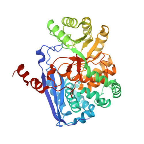
4A35

Crystal structure of human Mitochondrial enolase superfamily member 1 (ENOSF1)


Experimental Data Snapshot

  • Method: X-RAY DIFFRACTION
  • Resolution: 1.74 Å
  • R-Value Free: 0.176 
  • R-Value Work: 0.152 
  • R-Value Observed: 0.153 

wwPDB Validation   3D Report Full Report


This is version 1.3 of the entry. See complete history


Literature

Enzymatic and structural characterization of rTS gamma provides insights into the function of rTS beta.

Wichelecki, D.J.Froese, D.S.Kopec, J.Muniz, J.R.Yue, W.W.Gerlt, J.A.

(2014) Biochemistry 53: 2732-2738

  • DOI: https://doi.org/10.1021/bi500349e
  • Primary Citation of Related Structures:  
    4A35

  • PubMed Abstract: 

    In humans, the gene encoding a reverse thymidylate synthase (rTS) is transcribed in the reverse direction of the gene encoding thymidylate synthase (TS) that is involved in DNA biosynthesis. Three isoforms are found: α, β, and γ, with the transcript of the α-isoform overlapping with that of TS. rTSβ has been of interest since the discovery of its overexpression in methotrexate and 5-fluorouracil resistant cell lines. Despite more than 20 years of study, none of the rTS isoforms have been biochemically or structurally characterized. In this study, we identified rTSγ as an l-fuconate dehydratase and determined its high-resolution crystal structure. Our data provide an explanation for the observed difference in enzymatic activities between rTSβ and rTSγ, enabling more informed proposals for the possible function of rTSβ in chemotherapeutic resistance.


  • Organizational Affiliation

    Departments of Biochemistry and Chemistry, Institute for Genomic Biology, University of Illinois at Urbana-Champaign , Urbana, Illinois 61801, United States.


Macromolecules
Find similar proteins by:  (by identity cutoff)  |  3D Structure
Entity ID: 1
MoleculeChains Sequence LengthOrganismDetailsImage
MITOCHONDRIAL ENOLASE SUPERFAMILY MEMBER 1441Homo sapiensMutation(s): 0 
EC: 5 (PDB Primary Data), 4.2.1.68 (UniProt)
UniProt & NIH Common Fund Data Resources
Find proteins for Q7L5Y1 (Homo sapiens)
Explore Q7L5Y1 
Go to UniProtKB:  Q7L5Y1
PHAROS:  Q7L5Y1
GTEx:  ENSG00000132199 
Entity Groups  
Sequence Clusters30% Identity50% Identity70% Identity90% Identity95% Identity100% Identity
UniProt GroupQ7L5Y1
Sequence Annotations
Expand
  • Reference Sequence
Small Molecules
Ligands 2 Unique
IDChains Name / Formula / InChI Key2D Diagram3D Interactions
EDO
Query on EDO

Download Ideal Coordinates CCD File 
D [auth A]
E [auth A]
F [auth A]
G [auth A]
H [auth A]
D [auth A],
E [auth A],
F [auth A],
G [auth A],
H [auth A],
I [auth A],
J [auth A]
1,2-ETHANEDIOL
C2 H6 O2
LYCAIKOWRPUZTN-UHFFFAOYSA-N
MG
Query on MG

Download Ideal Coordinates CCD File 
B [auth A],
C [auth A]
MAGNESIUM ION
Mg
JLVVSXFLKOJNIY-UHFFFAOYSA-N
Experimental Data & Validation

Experimental Data

  • Method: X-RAY DIFFRACTION
  • Resolution: 1.74 Å
  • R-Value Free: 0.176 
  • R-Value Work: 0.152 
  • R-Value Observed: 0.153 
  • Space Group: P 65 2 2
Unit Cell:
Length ( Å )Angle ( ˚ )
a = 84.77α = 90
b = 84.77β = 90
c = 316.3γ = 120
Software Package:
Software NamePurpose
BUSTERrefinement
MOSFLMdata reduction
SCALEPACKdata scaling
SHARPphasing

Structure Validation

View Full Validation Report


Entry History 

Revision History  (Full details and data files)

  • Version 1.0: 2011-10-12
    Type: Initial release
  • Version 1.1: 2015-12-23
    Changes: Database references
  • Version 1.2: 2019-01-16
    Changes: Data collection, Database references
  • Version 1.3: 2024-05-08
    Changes: Data collection, Database references, Derived calculations, Other