4A2N | pdb_00004a2n

Crystal Structure of Ma-ICMT


Experimental Data Snapshot

  • Method: X-RAY DIFFRACTION
  • Resolution: 3.40 Å
  • R-Value Free: 
    0.275 (Depositor), 0.300 (DCC) 
  • R-Value Work: 
    0.238 (Depositor), 0.253 (DCC) 
  • R-Value Observed: 
    0.240 (Depositor) 

wwPDB Validation 3D Report Full Report

Validation slider image for 4A2N

Ligand Structure Quality Assessment 


This is version 1.3 of the entry. See complete history

Literature

Mechanism of Isoprenylcysteine Carboxyl Methylation from the Crystal Structure of the Integral Membrane Methyltransferase Icmt.

Yang, J.Kulkarni, K.Manolaridis, I.Zhang, Z.Dodd, R.B.Mas-Droux, C.Barford, D.

(2011) Mol Cell 44: 997

  • DOI: https://doi.org/10.1016/j.molcel.2011.10.020
  • Primary Citation Related Structures: 
    4A2N

  • PubMed Abstract: 

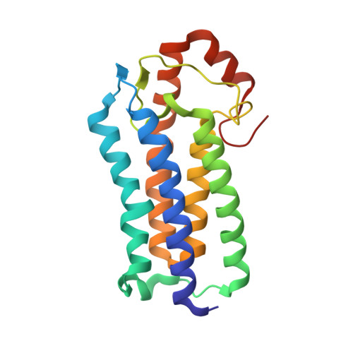
    The posttranslational modification of C-terminal CAAX motifs in proteins such as Ras, most Rho GTPases, and G protein γ subunits, plays an essential role in determining their subcellular localization and correct biological function. An integral membrane methyltransferase, isoprenylcysteine carboxyl methyltransferase (ICMT), catalyzes the final step of CAAX processing after prenylation of the cysteine residue and endoproteolysis of the -AAX motif. We have determined the crystal structure of a prokaryotic ICMT ortholog, revealing a markedly different architecture from conventional methyltransferases that utilize S-adenosyl-L-methionine (SAM) as a cofactor. ICMT comprises a core of five transmembrane α helices and a cofactor-binding pocket enclosed within a highly conserved C-terminal catalytic subdomain. A tunnel linking the reactive methyl group of SAM to the inner membrane provides access for the prenyl lipid substrate. This study explains how an integral membrane methyltransferase achieves recognition of both a hydrophilic cofactor and a lipophilic prenyl group attached to a polar protein substrate.


  • Organizational Affiliation
    • Division of Structural Biology, Institute of Cancer Research, Chester Beatty Laboratories, London SW3 6JB, UK.

Macromolecule Content 

  • Total Structure Weight: 24.94 kDa 
  • Atom Count: 1,612 
  • Modeled Residue Count: 192 
  • Deposited Residue Count: 194 
  • Unique protein chains: 1

Macromolecules

Find similar proteins by:|  3D Structure
Entity ID: 1
MoleculeChains  Sequence LengthOrganismDetailsImage
ISOPRENYLCYSTEINE CARBOXYL METHYLTRANSFERASEA [auth B]194Methanosarcina acetivoransMutation(s): 0 
EC: 2.1.1.100
Membrane Entity: Yes 
UniProt
Find proteins for Q8TMG0 (Methanosarcina acetivorans (strain ATCC 35395 / DSM 2834 / JCM 12185 / C2A))
Explore Q8TMG0 
Go to UniProtKB:  Q8TMG0
Entity Groups
Sequence Clusters30% Identity50% Identity70% Identity90% Identity95% Identity100% Identity
UniProt GroupQ8TMG0
Sequence Annotations
Expand
Reference Sequence

Experimental Data & Validation

Experimental Data

  • Method: X-RAY DIFFRACTION
  • Resolution: 3.40 Å
  • R-Value Free:  0.275 (Depositor), 0.300 (DCC) 
  • R-Value Work:  0.238 (Depositor), 0.253 (DCC) 
  • R-Value Observed: 0.240 (Depositor) 
Space Group: P 62 2 2
Unit Cell:
Length ( Å )Angle ( ˚ )
a = 114.92α = 90
b = 114.92β = 90
c = 169.291γ = 120
Software Package:
Software NamePurpose
MOSFLMdata reduction
SCALAdata scaling
PHENIXphasing
PHASERphasing
PHENIXrefinement

Structure Validation

View Full Validation Report


Ligand Structure Quality Assessment 


Entry History 

Deposition Data

Revision History  (Full details and data files)

  • Version 1.0: 2012-01-11
    Type: Initial release
  • Version 1.1: 2018-01-24
    Changes: Source and taxonomy
  • Version 1.2: 2024-05-08
    Changes: Data collection, Database references, Derived calculations, Other
  • Version 1.3: 2025-12-10
    Changes: Data collection, Structure summary