4QH7 | pdb_00004qh7

LC8 - Ana2 (159-168) Complex


Experimental Data Snapshot

  • Method: X-RAY DIFFRACTION
  • Resolution: 1.83 Å
  • R-Value Free: 
    0.207 (Depositor), 0.209 (DCC) 
  • R-Value Work: 
    0.176 (Depositor), 0.178 (DCC) 
  • R-Value Observed: 
    0.178 (Depositor) 

Starting Model: experimental
View more details

wwPDB Validation 3D Report Full Report

Validation slider image for 4QH7

This is version 1.2 of the entry. See complete history

Literature

The Mechanism of Dynein Light Chain LC8-mediated Oligomerization of the Ana2 Centriole Duplication Factor.

Slevin, L.K.Romes, E.M.Dandulakis, M.G.Slep, K.C.

(2014) J Biological Chem 289: 20727-20739

  • DOI: https://doi.org/10.1074/jbc.M114.576041
  • Primary Citation Related Structures: 
    4QH7, 4QH8

  • PubMed Abstract: 

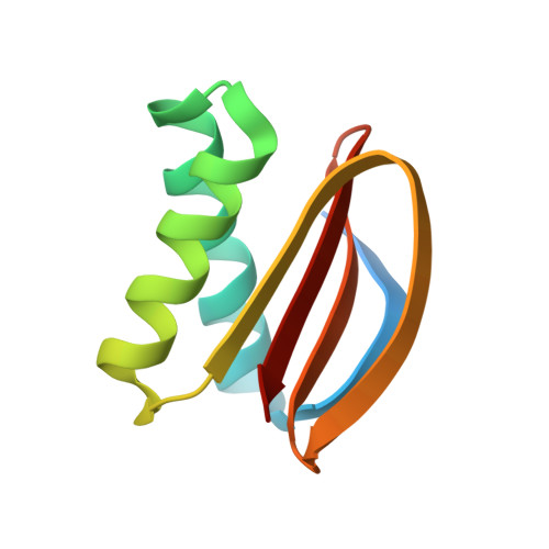
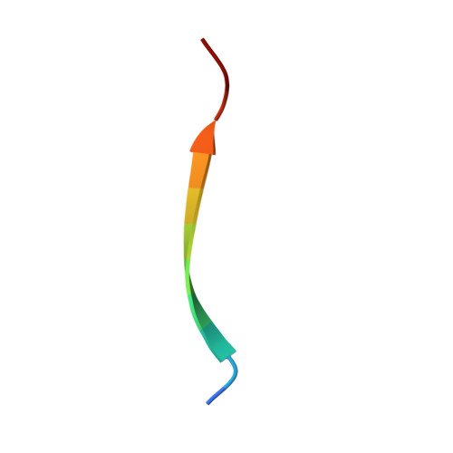
    Centrioles play a key role in nucleating polarized microtubule networks. In actively dividing cells, centrioles establish the bipolar mitotic spindle and are essential for genomic stability. Drosophila anastral spindle-2 (Ana2) is a conserved centriole duplication factor. Although recent work has demonstrated that an Ana2-dynein light chain (LC8) centriolar complex is critical for proper spindle positioning in neuroblasts, how Ana2 and LC8 interact is yet to be established. Here we examine the Ana2-LC8 interaction and map two LC8-binding sites within the central region of Ana2, Ana2M (residues 156-251). Ana2 LC8-binding site 1 contains a signature TQT motif and robustly binds LC8 (KD of 1.1 μm), whereas site 2 contains a TQC motif and binds LC8 with lower affinity (KD of 13 μm). Both LC8-binding sites flank a predicted ~34-residue α-helix. We present two independent atomic structures of LC8 dimers in complex with Ana2 LC8-binding site 1 and site 2 peptides. The Ana2 peptides form β-strands that extend a central composite LC8 β-sandwich. LC8 recognizes the signature TQT motif in the first LC8 binding site of Ana2, forming extensive van der Waals contacts and hydrogen bonding with the peptide, whereas the Ana2 site 2 TQC motif forms a uniquely extended β-strand, not observed in other dynein light chain-target complexes. Size exclusion chromatography coupled with multiangle static light scattering demonstrates that LC8 dimers bind Ana2M sites and induce Ana2 tetramerization, yielding an Ana2M4-LC88 complex. LC8-mediated Ana2 oligomerization probably enhances Ana2 avidity for centriole-binding factors and may bridge multiple factors as required during spindle positioning and centriole biogenesis.


  • Organizational Affiliation
    • From the Departments of Biology and.

Macromolecule Content 

  • Total Structure Weight: 48.33 kDa 
  • Atom Count: 3,356 
  • Modeled Residue Count: 376 
  • Deposited Residue Count: 424 
  • Unique protein chains: 2

Macromolecules

Find similar proteins by:|  3D Structure
Entity ID: 1
MoleculeChains  Sequence LengthOrganismDetailsImage
Dynein light chain 1, cytoplasmic
A, B, E, F
94Drosophila melanogasterMutation(s): 0 
Gene Names: ctpCdlc1ddlc1CG6998
UniProt
Find proteins for Q24117 (Drosophila melanogaster)
Explore Q24117 
Go to UniProtKB:  Q24117
Entity Groups
Sequence Clusters30% Identity50% Identity70% Identity90% Identity95% Identity100% Identity
UniProt GroupQ24117
Sequence Annotations
Expand
Reference Sequence
Find similar proteins by:  Sequence   |   3D Structure  
Entity ID: 2
MoleculeChains  Sequence LengthOrganismDetailsImage
Anastral spindle 2
C, D, G, H
12Drosophila melanogasterMutation(s): 0 
UniProt
Find proteins for Q9XZ31 (Drosophila melanogaster)
Explore Q9XZ31 
Go to UniProtKB:  Q9XZ31
Entity Groups
Sequence Clusters30% Identity50% Identity70% Identity90% Identity95% Identity100% Identity
UniProt GroupQ9XZ31
Sequence Annotations
Expand
Reference Sequence

Experimental Data & Validation

Experimental Data

  • Method: X-RAY DIFFRACTION
  • Resolution: 1.83 Å
  • R-Value Free:  0.207 (Depositor), 0.209 (DCC) 
  • R-Value Work:  0.176 (Depositor), 0.178 (DCC) 
  • R-Value Observed: 0.178 (Depositor) 
Space Group: P 21 21 21
Unit Cell:
Length ( Å )Angle ( ˚ )
a = 51.453α = 90
b = 77.913β = 90
c = 108.891γ = 90
Software Package:
Software NamePurpose
SERGUIdata collection
PHENIXmodel building
PHENIXrefinement
HKL-2000data reduction
HKL-2000data scaling
PHENIXphasing

Structure Validation

View Full Validation Report


Entry History 

Deposition Data

Revision History  (Full details and data files)

  • Version 1.0: 2014-06-11
    Type: Initial release
  • Version 1.1: 2014-08-20
    Changes: Database references
  • Version 1.2: 2023-09-20
    Changes: Data collection, Database references, Refinement description