4FBS | pdb_00004fbs

Structure of monomeric NT from Euprosthenops australis Major Ampullate Spidroin 1 (MaSp1)


Experimental Data Snapshot

  • Method: X-RAY DIFFRACTION
  • Resolution: 1.70 Å
  • R-Value Free: 
    0.196 (Depositor), 0.205 (DCC) 
  • R-Value Work: 
    0.142 (Depositor), 0.150 (DCC) 
  • R-Value Observed: 
    0.145 (Depositor) 

Starting Model: experimental
View more details

wwPDB Validation 3D Report Full Report

Validation slider image for 4FBS

This is version 1.2 of the entry. See complete history

Literature

pH-Dependent Dimerization of Spider Silk N-Terminal Domain Requires Relocation of a Wedged Tryptophan Side Chain

Jaudzems, K.Askarieh, G.Landreh, M.Nordling, K.Hedhammar, M.Jornvall, H.Rising, A.Knight, S.D.Johansson, J.

(2012) J Mol Biology 

  • DOI: https://doi.org/10.1016/j.jmb.2012.06.004
  • Primary Citation Related Structures: 
    2LPI, 2LPJ, 4FBS

  • PubMed Abstract: 

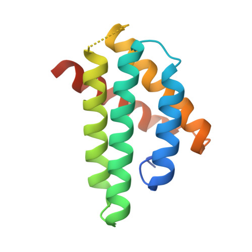
    Formation of spider silk from its constituent proteins-spidroins-involves changes from soluble helical/coil conformations to insoluble β-sheet aggregates. This conversion needs to be regulated to avoid precocious aggregation proximally in the silk gland while still allowing rapid silk assembly in the distal parts. Lowering of pH from about 7 to 6 is apparently important for silk formation. The spidroin N-terminal domain (NT) undergoes stable dimerization and structural changes in this pH region, but the underlying mechanisms are incompletely understood. Here, we determine the NMR and crystal structures of Euprosthenops australis NT mutated in the dimer interface (A72R). Also, the NMR structure of wild-type (wt) E. australis NT at pH7.2 and 300 mM sodium chloride was determined. The wt NT and A72R structures are monomers and virtually identical, but they differ from the subunit structure of dimeric wt NT mainly by having a tryptophan (W10) buried between helix 1 and helix 3, while W10 is surface exposed in the dimer. Wedging of the W10 side chain in monomeric NT tilts helix 3 approximately 5-6Å into a position that is incompatible with that of the observed dimer structure. The structural differences between monomeric and dimeric NT domains explain the tryptophan fluorescence patterns of NT at pH7 and pH6 and indicate that the biological function of NT depends on conversion between the two conformations.


  • Organizational Affiliation
    • Department of Physical Organic Chemistry, Latvian Institute of Organic Synthesis, Aizkraukles 21, Riga LV-1006, Latvia.

Macromolecule Content 

  • Total Structure Weight: 14.43 kDa 
  • Atom Count: 1,061 
  • Modeled Residue Count: 120 
  • Deposited Residue Count: 137 
  • Unique protein chains: 1

Macromolecules

Find similar proteins by:|  3D Structure
Entity ID: 1
MoleculeChains  Sequence LengthOrganismDetailsImage
Major ampullate spidroin 1137Euprosthenops australisMutation(s): 1 
Gene Names: MaSp1
UniProt
Find proteins for Q05H60 (Euprosthenops australis)
Explore Q05H60 
Go to UniProtKB:  Q05H60
Entity Groups
Sequence Clusters30% Identity50% Identity70% Identity90% Identity95% Identity100% Identity
UniProt GroupQ05H60
Sequence Annotations
Expand
Reference Sequence

Experimental Data & Validation

Experimental Data

  • Method: X-RAY DIFFRACTION
  • Resolution: 1.70 Å
  • R-Value Free:  0.196 (Depositor), 0.205 (DCC) 
  • R-Value Work:  0.142 (Depositor), 0.150 (DCC) 
  • R-Value Observed: 0.145 (Depositor) 
Space Group: P 1 21 1
Unit Cell:
Length ( Å )Angle ( ˚ )
a = 42.65α = 90
b = 33.244β = 115.37
c = 44.752γ = 90
Software Package:
Software NamePurpose
ADSCdata collection
AMoREphasing
PHENIXrefinement
MOSFLMdata reduction
SCALAdata scaling

Structure Validation

View Full Validation Report


Entry History 

Deposition Data

Revision History  (Full details and data files)

  • Version 1.0: 2012-08-01
    Type: Initial release
  • Version 1.1: 2024-03-20
    Changes: Data collection, Database references, Derived calculations
  • Version 1.2: 2024-04-03
    Changes: Refinement description