A sulfate pocket formed by three GoU pairs in the 0.97 A resolution X-ray structure of a nonameric RNA.
Masquida, B., Sauter, C., Westhof, E.(1999) RNA 5: 1384-1395
- PubMed: 10573129 Search on PubMedSearch on PubMed Central
- DOI: https://doi.org/10.1017/s1355838299991173
- Primary Citation Related Structures:
485D - PubMed Abstract:
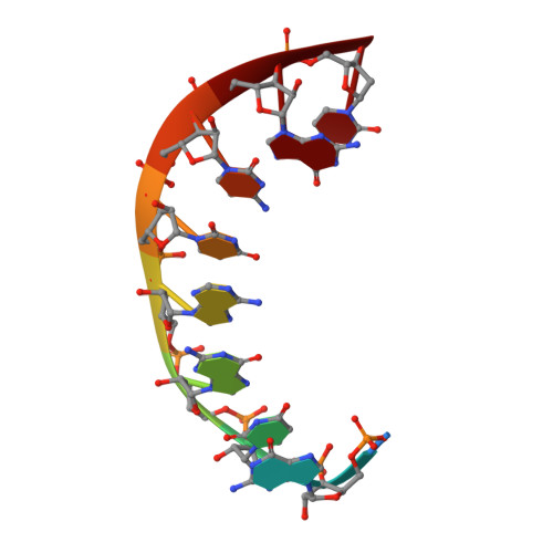
The crystal structure of the RNA duplex [r(CGUGAUCG)dC]2 has been solved at a resolution of 0.97 A. The model has been refined to R-work and R-free of 14.88% and 19.54% for 23,838 independent reflections. The base-pairing scheme forces the 5'-rC to be excluded from the helix and to be disordered. In the crystals, the sequence promotes the formation of two GoU wobble pairs that cluster around a crystallographic threefold axis in two different ways. In the first contact type, the GoU pairs are exclusively surrounded by water molecules, whereas in the other contact type, the three amino groups of the guanine residues of the symmetry-related GoU pairs trap a sulfate ion. This work provides the first example of the interaction of a GoU pair with a sulfate ion in a helical context. Despite the negative charge on the polynucleotide backbone, the guanine amino N2 is able to attract negatively charged groups that could, in the folding of complex RNA molecules, belong to a negative phosphodiester group from a neighboring strand and, in a RNA-protein complex, to a negative carboxyl group of an aspartate or glutamate side chain.
- Institut de Biologie Moléculaire et Cellulaire--Centre National de la Recherche Scientifique, UPR 9002 Structure des Macromolécules Biologiques et Mécanismes de Reconnaissance, Strasbourg, France.
Organizational Affiliation: