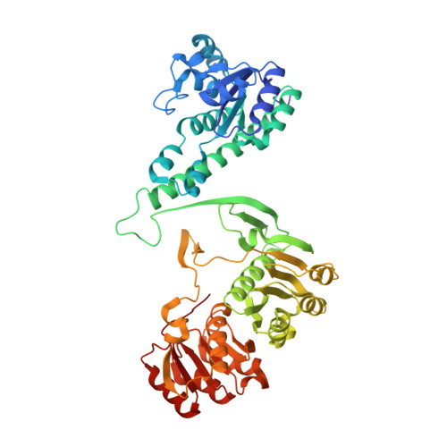
Structural Analyses of a Purine Biosynthetic Enzyme from Mycobacterium Tuberculosis Reveal a Novel Bound Nucleotide.
Le Nours, J., Bulloch, E.M.M., Zhang, Z., Greenwood, D.R., Middleditch, M.J., Dickson, J.M.J., Baker, E.N.(2011) J Biological Chem 286: 40706
- PubMed: 21956117
- DOI: https://doi.org/10.1074/jbc.M111.291138
- Primary Citation of Related Structures:
3ZZM, 4A1O - PubMed Abstract:
Enzymes of the de novo purine biosynthetic pathway have been identified as essential for the growth and survival of Mycobacterium tuberculosis and thus have potential for the development of anti-tuberculosis drugs. The final two steps of this pathway are carried out by the bifunctional enzyme 5-aminoimidazole-4-carboxamide ribonucleotide transformylase/inosine monophosphate cyclohydrolase (ATIC), also known as PurH. This enzyme has already been the target of anti-cancer drug development. We have determined the crystal structures of the M. tuberculosis ATIC (Rv0957) both with and without the substrate 5-aminoimidazole-4-carboxamide ribonucleotide, at resolutions of 2.5 and 2.2 Å, respectively. As for other ATIC enzymes, the protein is folded into two domains, the N-terminal domain (residues 1-212) containing the cyclohydrolase active site and the C-terminal domain (residues 222-523) containing the formyltransferase active site. An adventitiously bound nucleotide was found in the cyclohydrolase active site in both structures and was identified by NMR and mass spectral analysis as a novel 5-formyl derivative of an earlier intermediate in the biosynthetic pathway 4-carboxy-5-aminoimidazole ribonucleotide. This result and other studies suggest that this novel nucleotide is a cyclohydrolase inhibitor. The dimer formed by M. tuberculosis ATIC is different from those seen for human and avian ATICs, but it has a similar ∼50-Å separation of the two active sites of the bifunctional enzyme. Evidence in M. tuberculosis ATIC for reactivity of half-the-sites in the cyclohydrolase domains can be attributed to ligand-induced movements that propagate across the dimer interface and may be a common feature of ATIC enzymes.
- Maurice Wilkins Centre for Molecular Biodiscovery and School of Biological Sciences, University of Auckland, Private Bag 92019, Auckland, New Zealand.
Organizational Affiliation:


