3ZYW

Crystal structure of the first glutaredoxin domain of human glutaredoxin 3 (GLRX3)


Experimental Data Snapshot

  • Method: X-RAY DIFFRACTION
  • Resolution: 1.84 Å
  • R-Value Free: 0.250 
  • R-Value Work: 0.200 
  • R-Value Observed: 0.202 

wwPDB Validation   3D Report Full Report


This is version 1.2 of the entry. See complete history


Literature

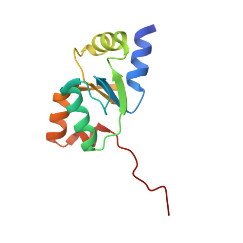
Crystal Structure of the First Glutaredoxin Domain of Human Glutaredoxin 3 (Glrx3)

Vollmar, M.Johansson, C.Cocking, R.Krojer, T.Muniz, J.R.C.Kavanagh, K.L.von Delft, F.Bountra, C.Arrowsmith, C.H.Weigelt, J.Edwards, A.Oppermann, U.

To be published.

Macromolecules
Find similar proteins by:  (by identity cutoff)  |  3D Structure
Entity ID: 1
MoleculeChains Sequence LengthOrganismDetailsImage
GLUTAREDOXIN-3
A, B
111Homo sapiensMutation(s): 0 
UniProt & NIH Common Fund Data Resources
Find proteins for O76003 (Homo sapiens)
Explore O76003 
Go to UniProtKB:  O76003
PHAROS:  O76003
GTEx:  ENSG00000108010 
Entity Groups  
Sequence Clusters30% Identity50% Identity70% Identity90% Identity95% Identity100% Identity
UniProt GroupO76003
Sequence Annotations
Expand
  • Reference Sequence
Experimental Data & Validation

Experimental Data

  • Method: X-RAY DIFFRACTION
  • Resolution: 1.84 Å
  • R-Value Free: 0.250 
  • R-Value Work: 0.200 
  • R-Value Observed: 0.202 
  • Space Group: P 2 21 21
Unit Cell:
Length ( Å )Angle ( ˚ )
a = 34.524α = 90
b = 87.052β = 90
c = 90.705γ = 90
Software Package:
Software NamePurpose
REFMACrefinement
XDSdata reduction
SCALAdata scaling
PHASERphasing

Structure Validation

View Full Validation Report


Entry History 

Revision History  (Full details and data files)

  • Version 1.0: 2012-02-29
    Type: Initial release
  • Version 1.1: 2018-01-24
    Changes: Database references, Structure summary
  • Version 1.2: 2023-12-20
    Changes: Data collection, Database references, Derived calculations, Other, Refinement description