


A Journey Through the Dap Kinase Architecture
De Diego, I., Kuper, J., Lehmann, F., Wilmanns, M.To be published.
Experimental Data Snapshot
Starting Model: experimental
View more details
Entity ID: 1 | |||||
|---|---|---|---|---|---|
| Molecule | Chains | Sequence Length | Organism | Details | Image |
| DEATH-ASSOCIATED PROTEIN KINASE 1 | 285 | Homo sapiens | Mutation(s): 0 EC: 2.7.11.1 | 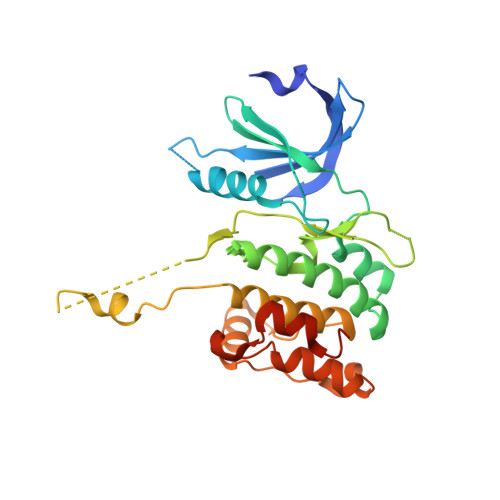 | |
UniProt & NIH Common Fund Data Resources | |||||
PHAROS: P53355 GTEx: ENSG00000196730 | |||||
Entity Groups | |||||
| Sequence Clusters | 30% Identity50% Identity70% Identity90% Identity95% Identity100% Identity | ||||
| UniProt Group | P53355 | ||||
Sequence AnnotationsExpand | |||||
Reference Sequence | |||||
| Ligands 2 Unique | |||||
|---|---|---|---|---|---|
| ID | Chains | Name / Formula / InChI Key | 2D Diagram | 3D Interactions | |
| ACP Download:Ideal Coordinates CCD File | E [auth A], J [auth B], L [auth C], M [auth D] | PHOSPHOMETHYLPHOSPHONIC ACID ADENYLATE ESTER C11 H18 N5 O12 P3 UFZTZBNSLXELAL-IOSLPCCCSA-N |  | ||
| MG Download:Ideal Coordinates CCD File | F [auth A] G [auth A] H [auth B] I [auth B] K [auth C] | MAGNESIUM ION Mg JLVVSXFLKOJNIY-UHFFFAOYSA-N |  | ||
| Length ( Å ) | Angle ( ˚ ) |
|---|---|
| a = 83.407 | α = 90 |
| b = 76.578 | β = 92.01 |
| c = 106.892 | γ = 90 |
| Software Name | Purpose |
|---|---|
| REFMAC | refinement |
| XDS | data reduction |
| SCALA | data scaling |
| PHASER | phasing |


