3ZVV | pdb_00003zvv

Fragment Bound to PI3Kinase gamma


Experimental Data Snapshot

  • Method: X-RAY DIFFRACTION
  • Resolution: 2.50 Å
  • R-Value Free: 
    0.242 (Depositor), 0.255 (DCC) 
  • R-Value Work: 
    0.192 (Depositor), 0.203 (DCC) 
  • R-Value Observed: 
    0.194 (Depositor) 

wwPDB Validation 3D Report Full Report

Validation slider image for 3ZVV

Ligand Structure Quality Assessment 


This is version 1.3 of the entry. See complete history

Literature

Fragment based discovery of a novel and selective PI3 kinase inhibitor.

Hughes, S.J.Millan, D.S.Kilty, I.C.Lewthwaite, R.A.Mathias, J.P.O'Reilly, M.A.Pannifer, A.Phelan, A.Stuhmeier, F.Baldock, D.A.Brown, D.G.

(2011) Bioorg Med Chem Lett 21: 6586-6590

  • DOI: https://doi.org/10.1016/j.bmcl.2011.07.117
  • Primary Citation Related Structures: 
    3ZVV, 3ZW3

  • PubMed Abstract: 

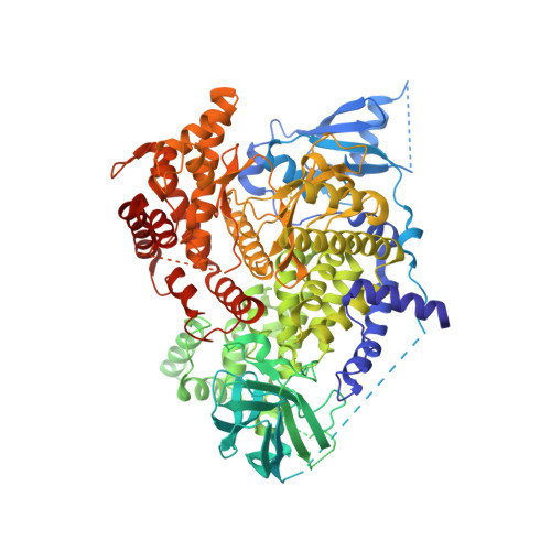
    We report the use of fragment screening and fragment based drug design to develop a PI3γ kinase fragment hit into a lead. Initial fragment hits were discovered by high concentration biochemical screening, followed by a round of virtual screening to identify additional ligand efficient fragments. These were developed into potent and ligand efficient lead compounds using structure guided fragment growing and merging strategies. This led to a potent, selective, and cell permeable PI3γ kinase inhibitor with good metabolic stability that was useful as a preclinical tool compound.


  • Organizational Affiliation
    • Worldwide Medicinal Chemistry, Pfizer Global Research and Development, Sandwich Laboratories, Ramsgate Road, Sandwich, Kent CT13 9NJ, UK. samantha.hughes@pfizer.com

Macromolecule Content 

  • Total Structure Weight: 110.89 kDa 
  • Atom Count: 7,023 
  • Modeled Residue Count: 829 
  • Deposited Residue Count: 966 
  • Unique protein chains: 1

Macromolecules

Find similar proteins by:|  3D Structure
Entity ID: 1
MoleculeChains  Sequence LengthOrganismDetailsImage
PHOSPHATIDYLINOSITOL-4,5-BISPHOSPHATE 3-KINASE CATALYTIC SUBUNIT GAMMA ISOFORM966Homo sapiensMutation(s): 0 
EC: 2.7.1.137 (PDB Primary Data), 2.7.1.153 (PDB Primary Data), 2.7.1.154 (UniProt), 2.7.11.1 (UniProt)
UniProt & NIH Common Fund Data Resources
Find proteins for P48736 (Homo sapiens)
Explore P48736 
Go to UniProtKB:  P48736
PHAROS:  P48736
GTEx:  ENSG00000105851 
Entity Groups
Sequence Clusters30% Identity50% Identity70% Identity90% Identity95% Identity100% Identity
UniProt GroupP48736
Sequence Annotations
Expand
Reference Sequence

Small Molecules

Ligands 1 Unique
IDChains Name / Formula / InChI Key2D Diagram3D Interactions
XAZ

Query on XAZ


Download:Ideal Coordinates CCD File
B [auth A]5,7-dimethylpyrazolo[1,5-a]pyrimidin-2-amine
C8 H10 N4
JTTOBYJUZIFSFJ-UHFFFAOYSA-N
Binding Affinity Annotations 
IDSourceBinding Affinity
XAZ BindingDB:  3ZVV IC50: 9.15e+5 (nM) from 1 assay(s)

Experimental Data & Validation

Experimental Data

  • Method: X-RAY DIFFRACTION
  • Resolution: 2.50 Å
  • R-Value Free:  0.242 (Depositor), 0.255 (DCC) 
  • R-Value Work:  0.192 (Depositor), 0.203 (DCC) 
  • R-Value Observed: 0.194 (Depositor) 
Space Group: C 1 2 1
Unit Cell:
Length ( Å )Angle ( ˚ )
a = 143.488α = 90
b = 68.031β = 95.16
c = 106.251γ = 90
Software Package:
Software NamePurpose
BUSTERrefinement
MOSFLMdata reduction
SCALAdata scaling

Structure Validation

View Full Validation Report


Ligand Structure Quality Assessment 


Entry History 

Deposition Data

Revision History  (Full details and data files)

  • Version 1.0: 2011-09-28
    Type: Initial release
  • Version 1.1: 2011-10-19
    Changes: Database references, Other
  • Version 1.2: 2018-02-07
    Changes: Database references, Source and taxonomy, Structure summary
  • Version 1.3: 2024-05-08
    Changes: Data collection, Database references, Derived calculations, Other