3ZRY | pdb_00003zry

Rotor architecture in the F(1)-c(10)-ring complex of the yeast F-ATP synthase


Experimental Data Snapshot

  • Method: X-RAY DIFFRACTION
  • Resolution: 6.50 Å
  • R-Value Free: 
    0.339 (Depositor), 0.346 (DCC) 
  • R-Value Work: 
    0.317 (Depositor), 0.306 (DCC) 
  • R-Value Observed: 
    0.318 (Depositor) 

Starting Model: experimental
View more details

wwPDB Validation 3D Report Full Report

Validation slider image for 3ZRY

Ligand Structure Quality Assessment 


This is version 2.1 of the entry. See complete history

Literature

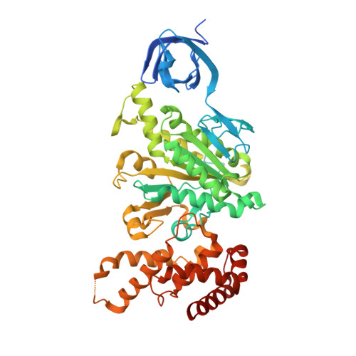
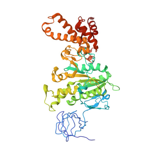
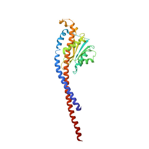
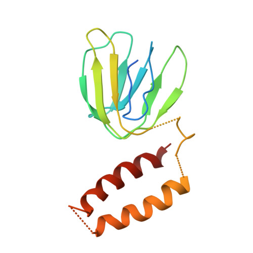
Rotor Architecture in the Yeast and Bovine F(1)-C-Ring Complexes of F-ATP Synthase.

Giraud, M.-F.Paumard, P.Sanchez, C.Brethes, D.Velours, J.Dautant, A.

(2012) J Struct Biol 177: 490

  • DOI: https://doi.org/10.1016/j.jsb.2011.10.015
  • Primary Citation Related Structures: 
    3ZRY

  • PubMed Abstract: 

    The F(1)F(O)-ATP synthase is a rotary molecular nanomotor. F(1) is a chemical motor driven by ATP hydrolysis while F(O) is an electrical motor driven by the proton flow. The two stepping motors are mechanically coupled through a common rotary shaft. Up to now, the three available crystal structures of the F(1)c(10) sub-complex of the yeast F(1)F(O)-ATP synthase were isomorphous and then named yF(1)c(10)(I). In this crystal form, significant interactions of the c(10)-ring with the F(1)-head of neighboring molecules affected the overall conformation of the F(1)-c-ring complex. The symmetry axis of the F(1)-head and the inertia axis of the c-ring were tilted near the interface between the F(1)-central stalk and the c-ring rotor, resulting in an unbalanced machine. We have solved a new crystal form of the F(1)c(10) complex, named yF(1)c(10)(II), inhibited by adenylyl-imidodiphosphate (AMP-PNP) and dicyclohexylcarbodiimide (DCCD), at 6.5Å resolution in which the crystal packing has a weaker influence over the conformation of the F(1)-c-ring complex. yF(1)c(10)(II) provides a model of a more efficient generator. yF(1)c(10)(II) and bovine bF(1)c(8) structures share a common rotor architecture with the inertia center of the F(1)-stator close to the rotor axis.


  • Organizational Affiliation
    • Univ. de Bordeaux, IBGC, UMR 5095, F-33000 Bordeaux, France. marie-france.giraud@ibgc.cnrs.fr

Macromolecule Content 

  • Total Structure Weight: 450.71 kDa 
  • Atom Count: 30,100 
  • Modeled Residue Count: 4,032 
  • Deposited Residue Count: 4,201 
  • Unique protein chains: 6

Macromolecules

Find similar proteins by:|  3D Structure
Entity ID: 1
MoleculeChains  Sequence LengthOrganismDetailsImage
ATP SYNTHASE SUBUNIT ALPHA, MITOCHONDRIAL
A, B, C
510Saccharomyces cerevisiaeMutation(s): 1 
Membrane Entity: Yes 
UniProt
Find proteins for P07251 (Saccharomyces cerevisiae (strain ATCC 204508 / S288c))
Explore P07251 
Go to UniProtKB:  P07251
Entity Groups
Sequence Clusters30% Identity50% Identity70% Identity90% Identity95% Identity100% Identity
UniProt GroupP07251
Sequence Annotations
Expand
Reference Sequence
Find similar proteins by:|  3D Structure
Entity ID: 2
MoleculeChains  Sequence LengthOrganismDetailsImage
ATP SYNTHASE SUBUNIT BETA, MITOCHONDRIAL
D, E, F
478Saccharomyces cerevisiaeMutation(s): 0 
EC: 3.6.3.14 (PDB Primary Data), 7.1.2.2 (UniProt)
Membrane Entity: Yes 
UniProt
Find proteins for P00830 (Saccharomyces cerevisiae (strain ATCC 204508 / S288c))
Explore P00830 
Go to UniProtKB:  P00830
Entity Groups
Sequence Clusters30% Identity50% Identity70% Identity90% Identity95% Identity100% Identity
UniProt GroupP00830
Sequence Annotations
Expand
Reference Sequence
Find similar proteins by:|  3D Structure
Entity ID: 3
MoleculeChains  Sequence LengthOrganismDetailsImage
ATP SYNTHASE SUBUNIT GAMMA, MITOCHONDRIAL278Saccharomyces cerevisiaeMutation(s): 0 
Membrane Entity: Yes 
UniProt
Find proteins for P38077 (Saccharomyces cerevisiae (strain ATCC 204508 / S288c))
Explore P38077 
Go to UniProtKB:  P38077
Entity Groups
Sequence Clusters30% Identity50% Identity70% Identity90% Identity95% Identity100% Identity
UniProt GroupP38077
Sequence Annotations
Expand
Reference Sequence
Find similar proteins by:|  3D Structure
Entity ID: 4
MoleculeChains  Sequence LengthOrganismDetailsImage
ATP SYNTHASE SUBUNIT DELTA, MITOCHONDRIAL138Saccharomyces cerevisiaeMutation(s): 0 
EC: 3.6.3.14
Membrane Entity: Yes 
UniProt
Find proteins for Q12165 (Saccharomyces cerevisiae (strain ATCC 204508 / S288c))
Explore Q12165 
Go to UniProtKB:  Q12165
Entity Groups
Sequence Clusters30% Identity50% Identity70% Identity90% Identity95% Identity100% Identity
UniProt GroupQ12165
Sequence Annotations
Expand
Reference Sequence
Find similar proteins by:|  3D Structure
Entity ID: 5
MoleculeChains  Sequence LengthOrganismDetailsImage
ATP SYNTHASE CATALYTIC SECTOR F1 EPSILON SUBUNIT61Saccharomyces cerevisiaeMutation(s): 0 
UniProt
Find proteins for E9P9X4 (Saccharomyces cerevisiae)
Explore E9P9X4 
Go to UniProtKB:  E9P9X4
Entity Groups
Sequence Clusters30% Identity50% Identity70% Identity90% Identity95% Identity100% Identity
UniProt GroupE9P9X4
Sequence Annotations
Expand
Reference Sequence
Find similar proteins by:|  3D Structure
Entity ID: 6
MoleculeChains  Sequence LengthOrganismDetailsImage
ATP SYNTHASE SUBUNIT 9, MITOCHONDRIAL
J, K, L, M, N
J, K, L, M, N, O, P, Q, R, S
76Saccharomyces cerevisiaeMutation(s): 0 
Membrane Entity: Yes 
UniProt
Find proteins for P61829 (Saccharomyces cerevisiae (strain ATCC 204508 / S288c))
Explore P61829 
Go to UniProtKB:  P61829
Entity Groups
Sequence Clusters30% Identity50% Identity70% Identity90% Identity95% Identity100% Identity
UniProt GroupP61829
Sequence Annotations
Expand
Reference Sequence

Small Molecules

Ligands 2 Unique
IDChains Name / Formula / InChI Key2D Diagram3D Interactions
ANP

Query on ANP


Download:Ideal Coordinates CCD File
BA [auth F],
T [auth A],
V [auth B],
X [auth C],
Z [auth D]
PHOSPHOAMINOPHOSPHONIC ACID-ADENYLATE ESTER
C10 H17 N6 O12 P3
PVKSNHVPLWYQGJ-KQYNXXCUSA-N
MG

Query on MG


Download:Ideal Coordinates CCD File
AA [auth D],
CA [auth F],
U [auth A],
W [auth B],
Y [auth C]
MAGNESIUM ION
Mg
JLVVSXFLKOJNIY-UHFFFAOYSA-N

Experimental Data & Validation

Experimental Data

  • Method: X-RAY DIFFRACTION
  • Resolution: 6.50 Å
  • R-Value Free:  0.339 (Depositor), 0.346 (DCC) 
  • R-Value Work:  0.317 (Depositor), 0.306 (DCC) 
  • R-Value Observed: 0.318 (Depositor) 
Space Group: P 1 21 1
Unit Cell:
Length ( Å )Angle ( ˚ )
a = 107.69α = 90
b = 174.91β = 93.92
c = 164.26γ = 90
Software Package:
Software NamePurpose
PHENIXrefinement
MOSFLMdata reduction
SCALAdata scaling
PHASERphasing

Structure Validation

View Full Validation Report


Ligand Structure Quality Assessment 


Entry History 

Deposition Data

Revision History  (Full details and data files)

  • Version 1.0: 2012-01-18
    Type: Initial release
  • Version 1.1: 2012-02-29
    Changes: Other
  • Version 2.0: 2019-01-23
    Changes: Advisory, Atomic model, Data collection, Other
  • Version 2.1: 2023-12-20
    Changes: Data collection, Database references, Derived calculations, Other, Refinement description