3ZP8 | pdb_00003zp8

HIGH-RESOLUTION FULL-LENGTH HAMMERHEAD RIBOZYME


Experimental Data Snapshot

  • Method: X-RAY DIFFRACTION
  • Resolution: 1.55 Å
  • R-Value Free: 
    0.216 (Depositor), 0.216 (DCC) 
  • R-Value Work: 
    0.187 (Depositor), 0.189 (DCC) 
  • R-Value Observed: 
    0.189 (Depositor) 

Starting Model: experimental
View more details

wwPDB Validation 3D Report Full Report

Validation slider image for 3ZP8

This is version 1.3 of the entry. See complete history

Literature

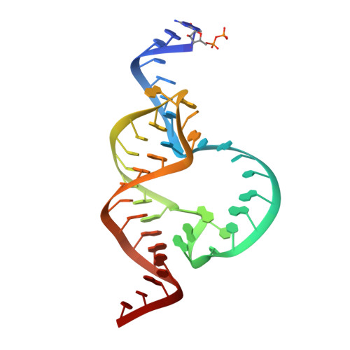
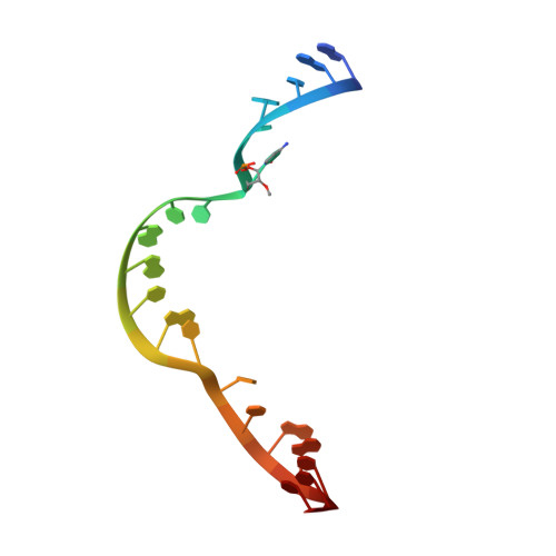
Active-Site Monovalent Cations Revealed in a 1.55 A Resolution Hammerhead Ribozyme Structure

Anderson, M.Schultz, E.Martick, M.Scott, W.G.

(2013) J Mol Biology 425: 3790

  • DOI: https://doi.org/10.1016/j.jmb.2013.05.017
  • Primary Citation Related Structures: 
    3ZP8

  • PubMed Abstract: 

    We have obtained a 1.55-Å crystal structure of a hammerhead ribozyme derived from Schistosoma mansoni under conditions that permit detailed observations of Na(+) ion binding in the ribozyme's active site. At least two such Na(+) ions are observed. The first Na(+) ion binds to the N7 of G10.1 and the adjacent A9 phosphate in a manner identical with that previously observed for divalent cations. A second Na(+) ion binds to the Hoogsteen face of G12, the general base in the hammerhead cleavage reaction, thereby potentially dissipating the negative charge of the catalytically active enolate form of the nucleotide base. A potential but more ambiguous third site bridges the A9 and scissile phosphates in a manner consistent with that of previous predictions. Hammerhead ribozymes have been observed to be active in the presence of high concentrations of monovalent cations, including Na(+), but the mechanism by which monovalent cations substitute for divalent cations in hammerhead catalysis remains unclear. Our results enable us to suggest that Na(+) directly and specifically substitutes for divalent cations in the hammerhead active site. The detailed geometry of the pre-catalytic active-site complex is also revealed with a new level of precision, thanks to the quality of the electron density maps obtained from what is currently the highest-resolution ribozyme structure in the Protein Data Bank.


  • Organizational Affiliation
    • Department of Chemistry and Biochemistry and The Center for the Molecular Biology of RNA, 228 Sinsheimer Laboratories, University of California at Santa Cruz, Santa Cruz, CA 95064, USA.

Macromolecule Content 

  • Total Structure Weight: 20.64 kDa 
  • Atom Count: 1,649 
  • Modeled Residue Count: 63 
  • Deposited Residue Count: 63 
  • Unique nucleic acid chains: 2

Macromolecules

Find similar nucleic acids by:  (by identity cutoff) 
Entity ID: 1
MoleculeChains LengthOrganismImage
HAMMERHEAD RIBOZYME, ENZYME STRAND43synthetic construct
Sequence Annotations
Expand
Reference Sequence
Find similar nucleic acids by:  Sequence
Entity ID: 2
MoleculeChains LengthOrganismImage
HAMMERHEAD RIBOZYME, SUBSTRATE STRAND20synthetic construct
Sequence Annotations
Expand
Reference Sequence

Small Molecules

Ligands 1 Unique
IDChains Name / Formula / InChI Key2D Diagram3D Interactions
NA

Query on NA


Download:Ideal Coordinates CCD File
C [auth A]
D [auth A]
E [auth A]
F [auth A]
G [auth A]
C [auth A],
D [auth A],
E [auth A],
F [auth A],
G [auth A],
H [auth A],
I [auth A],
J [auth A],
K [auth A],
L [auth A],
M [auth A],
N [auth A],
O [auth B],
P [auth B],
Q [auth B],
R [auth B]
SODIUM ION
Na
FKNQFGJONOIPTF-UHFFFAOYSA-N

Experimental Data & Validation

Experimental Data

  • Method: X-RAY DIFFRACTION
  • Resolution: 1.55 Å
  • R-Value Free:  0.216 (Depositor), 0.216 (DCC) 
  • R-Value Work:  0.187 (Depositor), 0.189 (DCC) 
  • R-Value Observed: 0.189 (Depositor) 
Space Group: C 1 2 1
Unit Cell:
Length ( Å )Angle ( ˚ )
a = 50.82α = 90
b = 68.53β = 112.35
c = 58.9γ = 90
Software Package:
Software NamePurpose
PHENIXrefinement
iMOSFLMdata reduction
PHENIXphasing

Structure Validation

View Full Validation Report


Entry History 

Deposition Data

Revision History  (Full details and data files)

  • Version 1.0: 2013-03-06
    Type: Initial release
  • Version 1.1: 2013-06-05
    Changes: Database references
  • Version 1.2: 2013-10-30
    Changes: Database references
  • Version 1.3: 2023-12-20
    Changes: Data collection, Database references, Derived calculations, Other, Refinement description