A Single Mutation in the Gste2 Gene Allows Tracking of Metabolically-Based Insecticide Resistance in a Major Malaria Vector.
Riveron, J.M., Yunta, C., Ibrahim, S.S., Djouaka, R., Irving, H., Menze, B.D., Ismail, H.M., Hemingway, J., Ranson, H., Albert, A., Wondji, C.S.(2014) Genome Biology 15: R27
- PubMed: 24565444 Search on PubMedSearch on PubMed Central
- DOI: https://doi.org/10.1186/gb-2014-15-2-r27
- Primary Citation Related Structures:
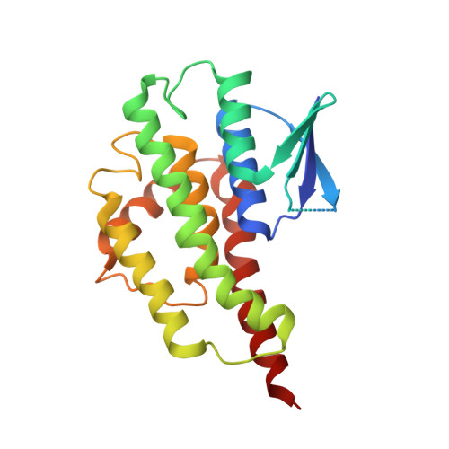
3ZMK, 3ZML - PubMed Abstract:
Metabolic resistance to insecticides is the biggest threat to the continued effectiveness of malaria vector control. However, its underlying molecular basis, crucial for successful resistance management, remains poorly characterized. Here, we demonstrate that the single amino acid change L119F in an upregulated glutathione S-transferase gene, GSTe2, confers high levels of metabolic resistance to DDT in the malaria vector Anopheles funestus. Genome-wide transcription analysis revealed that GSTe2 was the most over-expressed detoxification gene in DDT and permethrin-resistant mosquitoes from Benin. Transgenic expression of GSTe2 in Drosophila melanogaster demonstrated that over-transcription of this gene alone confers DDT resistance and cross-resistance to pyrethroids. Analysis of GSTe2 polymorphism established that the point mutation is tightly associated with metabolic resistance to DDT and its geographical distribution strongly correlates with DDT resistance patterns across Africa. Functional characterization of recombinant GSTe2 further supports the role of the L119F mutation, with the resistant allele being more efficient at metabolizing DDT than the susceptible one. Importantly, we also show that GSTe2 directly metabolizes the pyrethroid permethrin. Structural analysis reveals that the mutation confers resistance by enlarging the GSTe2 DDT-binding cavity, leading to increased DDT access and metabolism. Furthermore, we show that GSTe2 is under strong directional selection in resistant populations, and a restriction of gene flow is observed between African regions, enabling the prediction of the future spread of this resistance. This first DNA-based metabolic resistance marker in mosquitoes provides an essential tool to track the evolution of resistance and to design suitable resistance management strategies.