3ZKE

Structure of LC8 in complex with Nek9 peptide


Experimental Data Snapshot

  • Method: X-RAY DIFFRACTION
  • Resolution: 2.20 Å
  • R-Value Free: 0.228 
  • R-Value Work: 0.196 
  • R-Value Observed: 0.197 

wwPDB Validation   3D Report Full Report


This is version 1.3 of the entry. See complete history


Literature

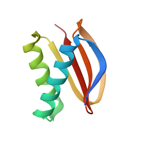
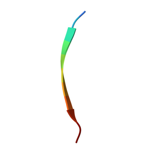
Structural Analysis of the Regulation of the Dynll/Lc8 Binding to Nek9 by Phosphorylation

Gallego, P.Velazquez-Campoy, A.Regue, L.Roig, J.Reverter, D.

(2013) J Biol Chem 288: 12283

  • DOI: https://doi.org/10.1074/jbc.M113.459149
  • Primary Citation of Related Structures:  
    3ZKE, 3ZKF

  • PubMed Abstract: 

    The NIMA family protein kinases Nek9/Nercc1, Nek6, and Nek7 constitute a signaling module activated in early mitosis involved in the control of spindle organization. DYNLL/LC8 (dynein light chain 8) was originally described as a component of the dynein complex, but the recent discovery of multiple interaction partners for LC8 has suggested that it has a general role as a dimerization hub that organizes different protein partners. Recent experiments suggested that LC8 binding to Nek9 was regulated by Nek9 autophosphorylation on Ser(944), a residue immediately located N-terminal to the LC8 conserved (K/R)xTQT binding motif, and that this was crucial for the control of signal transduction through the Nek/Nek6/7 module. In the present work, we present two crystal structures of LC8 with a peptide corresponding to the Nek9 binding region with and without a phosphorylation on Ser(944). Structural analysis of LC8 with both Nek9 peptides, together with different biophysical experiments, explains the observed diminished binding affinity of Nek9 to LC8 upon phosphorylation on Ser(944) within the Nek9 sequence, thus shedding light into a novel phosphorylation regulatory mechanism that interferes with LC8 protein · protein complex formation.


  • Organizational Affiliation

    Structural Biology Unit, Institut de Biotecnologia i Biomedicina, Universitat Autònoma de Barcelona, 08193 Bellaterra, Spain.


Macromolecules
Find similar proteins by:  (by identity cutoff)  |  3D Structure
Entity ID: 1
MoleculeChains Sequence LengthOrganismDetailsImage
DYNEIN LIGHT CHAIN 1, CYTOPLASMIC
A, C, E, G, I
A, C, E, G, I, K
89Homo sapiensMutation(s): 0 
UniProt & NIH Common Fund Data Resources
Find proteins for P63167 (Homo sapiens)
Explore P63167 
Go to UniProtKB:  P63167
PHAROS:  P63167
GTEx:  ENSG00000088986 
Entity Groups  
Sequence Clusters30% Identity50% Identity70% Identity90% Identity95% Identity100% Identity
UniProt GroupP63167
Sequence Annotations
Expand
  • Reference Sequence

Find similar proteins by:  Sequence   |   3D Structure  

Entity ID: 2
MoleculeChains Sequence LengthOrganismDetailsImage
NEK9 PROTEIN
B, D, F, H, J
B, D, F, H, J, L
11Homo sapiensMutation(s): 0 
UniProt & NIH Common Fund Data Resources
Find proteins for Q8TD19 (Homo sapiens)
Explore Q8TD19 
Go to UniProtKB:  Q8TD19
PHAROS:  Q8TD19
GTEx:  ENSG00000119638 
Entity Groups  
Sequence Clusters30% Identity50% Identity70% Identity90% Identity95% Identity100% Identity
UniProt GroupQ8TD19
Sequence Annotations
Expand
  • Reference Sequence
Experimental Data & Validation

Experimental Data

  • Method: X-RAY DIFFRACTION
  • Resolution: 2.20 Å
  • R-Value Free: 0.228 
  • R-Value Work: 0.196 
  • R-Value Observed: 0.197 
  • Space Group: P 21 21 21
Unit Cell:
Length ( Å )Angle ( ˚ )
a = 39.984α = 90
b = 105.124β = 90
c = 133.899γ = 90
Software Package:
Software NamePurpose
REFMACrefinement
XDSdata reduction
SCALAdata scaling
MOLREPphasing

Structure Validation

View Full Validation Report


Entry History 

Deposition Data

Revision History  (Full details and data files)

  • Version 1.0: 2013-03-20
    Type: Initial release
  • Version 1.1: 2013-04-03
    Changes: Database references
  • Version 1.2: 2013-05-15
    Changes: Database references
  • Version 1.3: 2013-05-22
    Changes: Derived calculations