3ZIR | pdb_00003zir

minor-site specific NLS (B141)


Experimental Data Snapshot

  • Method: X-RAY DIFFRACTION
  • Resolution: 2.30 Å
  • R-Value Free: 
    0.202 (Depositor), 0.221 (DCC) 
  • R-Value Work: 
    0.175 (Depositor), 0.195 (DCC) 
  • R-Value Observed: 
    0.177 (Depositor) 

Starting Model: experimental
View more details

wwPDB Validation 3D Report Full Report

Validation slider image for 3ZIR

This is version 1.2 of the entry. See complete history

Literature

Distinctive Conformation of Minor Site-Specific Nuclear Localization Signals Bound to Importin-Alpha

Chang, C.-W.Counago, R.M.Williams, S.J.Boden, M.Kobe, B.

(2013) Traffic 14: 1144

  • DOI: https://doi.org/10.1111/tra.12098
  • Primary Citation Related Structures: 
    3ZIN, 3ZIO, 3ZIP, 3ZIQ, 3ZIR

  • PubMed Abstract: 

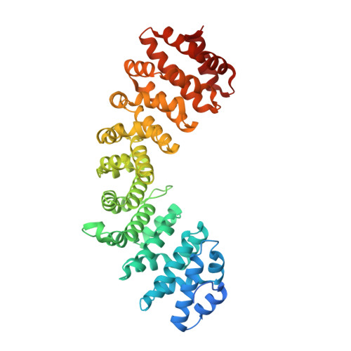
    Nuclear localization signals (NLSs) contain one or two clusters of basic residues and are recognized by the import receptor importin-α. There are two NLS-binding sites (major and minor) on importin-α and the major NLS-binding site is considered to be the primary binding site. Here, we used crystallographic and biochemical methods to investigate the binding between importin-α and predicted 'minor site-specific' NLSs: four peptide library-derived peptides, and the NLS from mouse RNA helicase II/Guα. The crystal structures reveal that these atypical NLSs indeed preferentially bind to the minor NLS-binding site. Unlike previously characterized NLSs, the C-terminal residues of these NLSs form an α-helical turn, stabilized by internal H-bond and cation-π interactions between the aromatic residues from the NLSs and the positively charged residues from importin-α. This helical turn sterically hinders binding at the major NLS-binding site, explaining the minor-site preference. Our data suggest the sequence RXXKR[K/X][F/Y/W]XXAF as the optimal minor NLS-binding site-specific motif, which may help identify novel proteins with atypical NLSs.


  • Organizational Affiliation
    • School of Chemistry and Molecular Biosciences and Institute for Molecular Bioscience, University of Queensland, Brisbane, Qld, 4072, Australia; Australian Infectious Diseases Research Centre, University of Queensland, Brisbane, Qld, 4072, Australia.

Macromolecule Content 

  • Total Structure Weight: 52.87 kDa 
  • Atom Count: 3,561 
  • Modeled Residue Count: 443 
  • Deposited Residue Count: 483 
  • Unique protein chains: 2

Macromolecules

Find similar proteins by:|  3D Structure
Entity ID: 1
MoleculeChains  Sequence LengthOrganismDetailsImage
IMPORTIN SUBUNIT ALPHA-2461Mus musculusMutation(s): 0 
UniProt & NIH Common Fund Data Resources
Find proteins for P52293 (Mus musculus)
Explore P52293 
Go to UniProtKB:  P52293
IMPC:  MGI:103561
Entity Groups
Sequence Clusters30% Identity50% Identity70% Identity90% Identity95% Identity100% Identity
UniProt GroupP52293
Sequence Annotations
Expand
Reference Sequence
Find similar proteins by:  Sequence   |   3D Structure  
Entity ID: 2
MoleculeChains  Sequence LengthOrganismDetailsImage
B141NLS
B, C
11synthetic constructMutation(s): 0 

Experimental Data & Validation

Experimental Data

  • Method: X-RAY DIFFRACTION
  • Resolution: 2.30 Å
  • R-Value Free:  0.202 (Depositor), 0.221 (DCC) 
  • R-Value Work:  0.175 (Depositor), 0.195 (DCC) 
  • R-Value Observed: 0.177 (Depositor) 
Space Group: P 21 21 21
Unit Cell:
Length ( Å )Angle ( ˚ )
a = 79α = 90
b = 90.34β = 90
c = 100.84γ = 90
Software Package:
Software NamePurpose
BUSTERrefinement
XDSdata reduction
SCALAdata scaling
MOLREPphasing

Structure Validation

View Full Validation Report


Entry History 

Deposition Data

Revision History  (Full details and data files)

  • Version 1.0: 2013-08-21
    Type: Initial release
  • Version 1.1: 2013-10-30
    Changes: Database references
  • Version 1.2: 2023-12-20
    Changes: Data collection, Database references, Other, Refinement description