3ZHN | pdb_00003zhn

Crystal structure of the T6SS lipoprotein TssJ1 from Pseudomonas aeruginosa


Experimental Data Snapshot

  • Method: X-RAY DIFFRACTION
  • Resolution: 1.40 Å
  • R-Value Free: 
    0.202 (Depositor), 0.210 (DCC) 
  • R-Value Work: 
    0.161 (Depositor) 
  • R-Value Observed: 
    0.163 (Depositor) 

wwPDB Validation 3D Report Full Report

Validation slider image for 3ZHN

This is version 2.0 of the entry. See complete history

Literature

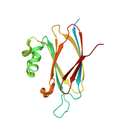
Structure of the T6Ss Lipoprotein Tssj1 from Pseudomonas Aeruginosa.

Robb, C.S.Assmus, M.Nano, F.E.Boraston, A.B.

(2013) Acta Crystallogr Sect F Struct Biol Cryst Commun 69: 607

  • DOI: https://doi.org/10.1107/S1744309113012220
  • Primary Citation Related Structures: 
    3ZHN

  • PubMed Abstract: 

    The type VI secretion system of Pseudomonas aeruginosa has been shown to be responsible for the translocation of bacteriolytic effectors into competing bacteria. A mechanistic understanding of this widely distributed secretion system is developing and structural studies of its components are ongoing. Two representative structures of one highly conserved component, TssJ, from Escherichia coli and Serratia marcescens have been published. Here, the X-ray crystal structure of TssJ1 from P. aeruginosa is presented at 1.4 Å resolution. The overall structure is conserved among the three proteins. This finding suggests that the homologues function in a similar manner and bolsters the understanding of the structure of this family of proteins.


  • Organizational Affiliation
    • Department of Biochemistry and Microbiology, University of Victoria, PO Box 3055 STN CSC, Victoria, British Columbia V8W 3P6, Canada.

Macromolecule Content 

  • Total Structure Weight: 19.09 kDa 
  • Atom Count: 1,171 
  • Modeled Residue Count: 131 
  • Deposited Residue Count: 159 
  • Unique protein chains: 1

Macromolecules

Find similar proteins by:|  3D Structure
Entity ID: 1
MoleculeChains  Sequence LengthOrganismDetailsImage
PA_0080159Pseudomonas aeruginosa PAO1Mutation(s): 0 
UniProt
Find proteins for Q9I752 (Pseudomonas aeruginosa (strain ATCC 15692 / DSM 22644 / CIP 104116 / JCM 14847 / LMG 12228 / 1C / PRS 101 / PAO1))
Explore Q9I752 
Go to UniProtKB:  Q9I752
Entity Groups
Sequence Clusters30% Identity50% Identity70% Identity90% Identity95% Identity100% Identity
UniProt GroupQ9I752
Sequence Annotations
Expand
Reference Sequence

Small Molecules

Ligands 1 Unique
IDChains Name / Formula / InChI Key2D Diagram3D Interactions
IOD

Query on IOD


Download:Ideal Coordinates CCD File
B [auth A]
C [auth A]
D [auth A]
E [auth A]
F [auth A]
B [auth A],
C [auth A],
D [auth A],
E [auth A],
F [auth A],
G [auth A],
H [auth A],
I [auth A],
J [auth A],
K [auth A],
L [auth A],
M [auth A]
IODIDE ION
I
XMBWDFGMSWQBCA-UHFFFAOYSA-M

Experimental Data & Validation

Experimental Data

  • Method: X-RAY DIFFRACTION
  • Resolution: 1.40 Å
  • R-Value Free:  0.202 (Depositor), 0.210 (DCC) 
  • R-Value Work:  0.161 (Depositor) 
  • R-Value Observed: 0.163 (Depositor) 
Space Group: P 2 21 21
Unit Cell:
Length ( Å )Angle ( ˚ )
a = 38.315α = 90
b = 41.637β = 90
c = 76.947γ = 90
Software Package:
Software NamePurpose
d*TREKdata reduction
SCALEPACKdata scaling
SHELXphasing
PHASERphasing
REFMACrefinement

Structure Validation

View Full Validation Report


Entry History 

Deposition Data

Revision History  (Full details and data files)

  • Version 1.0: 2013-06-12
    Type: Initial release
  • Version 2.0: 2024-05-08
    Changes: Atomic model, Data collection, Database references, Derived calculations, Other