3ZGI | pdb_00003zgi

Crystal structure of the KRT10-binding region domain of the pneumococcal serine rich repeat protein PsrP


Experimental Data Snapshot

  • Method: X-RAY DIFFRACTION
  • Resolution: 2.25 Å
  • R-Value Free: 
    0.214 (Depositor), 0.221 (DCC) 
  • R-Value Work: 
    0.186 (Depositor), 0.191 (DCC) 
  • R-Value Observed: 
    0.187 (Depositor) 

wwPDB Validation 3D Report Full Report

Validation slider image for 3ZGI

This is version 1.3 of the entry. See complete history

Literature

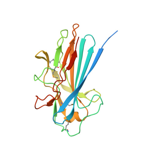
The Basic Keratin 10-Binding Domain of the Virulence-Associated Pneumococcal Serine-Rich Protein Psrp Adopts a Novel Mscramm Fold.

Schulte, T.Lofling, J.Mikaelsson, C.Kikhney, A.Hentrich, K.Diamante, A.Ebel, C.Normark, S.Svergun, D.Henriques-Normark, B.Achour, A.

(2014) Open Biol 4: 0090

  • DOI: https://doi.org/10.1098/rsob.130090
  • Primary Citation Related Structures: 
    3ZGH, 3ZGI

  • PubMed Abstract: 

    Streptococcus pneumoniae is a major human pathogen, and a leading cause of disease and death worldwide. Pneumococcal invasive disease is triggered by initial asymptomatic colonization of the human upper respiratory tract. The pneumococcal serine-rich repeat protein (PsrP) is a lung-specific virulence factor whose functional binding region (BR) binds to keratin-10 (KRT10) and promotes pneumococcal biofilm formation through self-oligomerization. We present the crystal structure of the KRT10-binding domain of PsrP (BR187-385) determined to 2.0 Å resolution. BR187-385 adopts a novel variant of the DEv-IgG fold, typical for microbial surface components recognizing adhesive matrix molecules adhesins, despite very low sequence identity. An extended β-sheet on one side of the compressed, two-sided barrel presents a basic groove that possibly binds to the acidic helical rod domain of KRT10. Our study also demonstrates the importance of the other side of the barrel, formed by extensive well-ordered loops and stabilized by short β-strands, for interaction with KRT10.


  • Organizational Affiliation
    • Science for Life Laboratory, Center for Infectious Medicine (CIM), Department of Medicine, Karolinska University Hospital Huddinge, Karolinska Institutet Science Park, Tomtebodavägen 23A Solna, Stockholm 17165, Sweden.

Macromolecule Content 

  • Total Structure Weight: 67.62 kDa 
  • Atom Count: 4,171 
  • Modeled Residue Count: 522 
  • Deposited Residue Count: 615 
  • Unique protein chains: 1

Macromolecules

Find similar proteins by:|  3D Structure
Entity ID: 1
MoleculeChains  Sequence LengthOrganismDetailsImage
CELL WALL SURFACE ANCHOR FAMILY PROTEIN
A, B, C
205Streptococcus pneumoniae TIGR4Mutation(s): 0 
UniProt
Find proteins for A0A0H2URK1 (Streptococcus pneumoniae serotype 4 (strain ATCC BAA-334 / TIGR4))
Explore A0A0H2URK1 
Go to UniProtKB:  A0A0H2URK1
Entity Groups
Sequence Clusters30% Identity50% Identity70% Identity90% Identity95% Identity100% Identity
UniProt GroupA0A0H2URK1
Sequence Annotations
Expand
Reference Sequence

Small Molecules

Modified Residues  1 Unique
IDChains TypeFormula2D DiagramParent
MSE
Query on MSE
A, B, C
L-PEPTIDE LINKINGC5 H11 N O2 SeMET

Experimental Data & Validation

Experimental Data

  • Method: X-RAY DIFFRACTION
  • Resolution: 2.25 Å
  • R-Value Free:  0.214 (Depositor), 0.221 (DCC) 
  • R-Value Work:  0.186 (Depositor), 0.191 (DCC) 
  • R-Value Observed: 0.187 (Depositor) 
Space Group: P 41 21 2
Unit Cell:
Length ( Å )Angle ( ˚ )
a = 105.58α = 90
b = 105.58β = 90
c = 120.45γ = 90
Software Package:
Software NamePurpose
PHENIXrefinement
XDSdata reduction
XSCALEdata scaling
Auto-Rickshawphasing

Structure Validation

View Full Validation Report


Entry History 

Revision History  (Full details and data files)

  • Version 1.0: 2013-12-25
    Type: Initial release
  • Version 1.1: 2014-01-29
    Changes: Database references
  • Version 1.2: 2019-05-08
    Changes: Data collection, Derived calculations, Experimental preparation, Other
  • Version 1.3: 2024-10-16
    Changes: Data collection, Database references, Derived calculations, Other, Structure summary