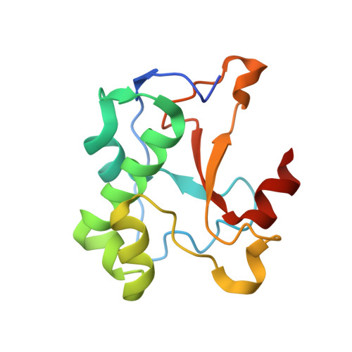
3ZFK

N-terminal truncated Nuclease Domain of Colicin E7


Experimental Data Snapshot

  • Method: X-RAY DIFFRACTION
  • Resolution: 1.70 Å
  • R-Value Free: 0.234 
  • R-Value Work: 0.200 
  • R-Value Observed: 0.202 

Starting Model: experimental
View more details

wwPDB Validation   3D Report Full Report


This is version 3.0 of the entry. See complete history


Literature

A New Insight Into the Zinc-Dependent DNA-Cleavage by the Colicin E7 Nuclease: A Crystallographic and Computational Study.

Czene, A.Toth, E.Nemeth, E.Otten, H.Poulsen, J.N.Christensen, H.E.M.Rulisek, L.Nagata, K.Larsen, S.Gyurcsik, B.

(2014) Metallomics 6: 2090

  • DOI: https://doi.org/10.1039/c4mt00195h
  • Primary Citation of Related Structures:  
    3ZFK

  • PubMed Abstract: 

    The nuclease domain of colicin E7 metallonuclease (NColE7) contains its active centre at the C-terminus. The mutant ΔN4-NColE7-C* - where the four N-terminal residues including the positively charged K446, R447 and K449 are replaced with eight residues from the GST tag - is catalytically inactive. The crystal structure of this mutant demonstrates that its overall fold is very similar to that of the native NColE7 structure. This implicates the stabilizing effect of the remaining N-terminal sequence on the structure of the C-terminal catalytic site and the essential role of the deleted residues in the mechanism of the catalyzed reaction. Complementary QM/MM calculations on the protein-DNA complexes support the less favourable cleavage by the mutant protein than by NColE7. Furthermore, a water molecule as a possible ligand for the Zn(2+)-ion is proposed to play a role in the catalytic process. These results suggest that the mechanism of the Zn(2+)-containing HNH nucleases needs to be further studied and discussed.


  • Organizational Affiliation

    MTA-SZTE Bioinorganic Chemistry Research Group, Dóm tér 7, H-6720 Szeged, Hungary.


Macromolecules
Find similar proteins by:  (by identity cutoff)  |  3D Structure
Entity ID: 1
MoleculeChains Sequence LengthOrganismDetailsImage
COLICIN-E7
A, B
135Escherichia coli BL21(DE3)Mutation(s): 0 
EC: 3.1
UniProt
Find proteins for Q47112 (Escherichia coli)
Explore Q47112 
Go to UniProtKB:  Q47112
Entity Groups  
Sequence Clusters30% Identity50% Identity70% Identity90% Identity95% Identity100% Identity
UniProt GroupQ47112
Sequence Annotations
Expand
  • Reference Sequence
Small Molecules
Ligands 4 Unique
IDChains Name / Formula / InChI Key2D Diagram3D Interactions
SO4
Query on SO4

Download Ideal Coordinates CCD File 
E [auth A]
F [auth A]
G [auth A]
H [auth A]
I [auth A]
E [auth A],
F [auth A],
G [auth A],
H [auth A],
I [auth A],
L [auth B],
M [auth B],
N [auth B],
O [auth B]
SULFATE ION
O4 S
QAOWNCQODCNURD-UHFFFAOYSA-L
ZN
Query on ZN

Download Ideal Coordinates CCD File 
C [auth A],
J [auth B]
ZINC ION
Zn
PTFCDOFLOPIGGS-UHFFFAOYSA-N
ACT
Query on ACT

Download Ideal Coordinates CCD File 
P [auth B],
Q [auth B]
ACETATE ION
C2 H3 O2
QTBSBXVTEAMEQO-UHFFFAOYSA-M
CL
Query on CL

Download Ideal Coordinates CCD File 
D [auth A],
K [auth B]
CHLORIDE ION
Cl
VEXZGXHMUGYJMC-UHFFFAOYSA-M
Experimental Data & Validation

Experimental Data

  • Method: X-RAY DIFFRACTION
  • Resolution: 1.70 Å
  • R-Value Free: 0.234 
  • R-Value Work: 0.200 
  • R-Value Observed: 0.202 
  • Space Group: P 32
Unit Cell:
Length ( Å )Angle ( ˚ )
a = 55.4α = 90
b = 55.4β = 90
c = 73.2γ = 120
Software Package:
Software NamePurpose
PHENIXrefinement
XDSdata reduction
XSCALEdata scaling
MOLREPphasing

Structure Validation

View Full Validation Report


Entry History 

Deposition Data

Revision History  (Full details and data files)

  • Version 1.0: 2013-12-18
    Type: Initial release
  • Version 1.1: 2014-08-13
    Changes: Atomic model, Derived calculations, Other
  • Version 1.2: 2014-12-24
    Changes: Database references
  • Version 2.0: 2018-01-17
    Changes: Atomic model, Data collection
  • Version 3.0: 2023-12-20
    Changes: Atomic model, Data collection, Database references, Derived calculations, Other, Refinement description