3ZDO | pdb_00003zdo

Tetramerization domain of Measles virus phosphoprotein


Experimental Data Snapshot

  • Method: X-RAY DIFFRACTION
  • Resolution: 2.07 Å
  • R-Value Free: 
    0.284 (Depositor), 0.283 (DCC) 
  • R-Value Work: 
    0.210 (Depositor), 0.210 (DCC) 
  • R-Value Observed: 
    0.213 (Depositor) 

Starting Model: in silico
View more details

wwPDB Validation 3D Report Full Report

Validation slider image for 3ZDO

This is version 1.2 of the entry. See complete history

Literature

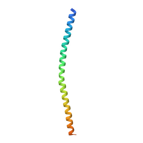
Structure of the Tetramerization Domain of Measles Virus Phosphoprotein.

Communie, G.Crepin, T.Maurin, D.Ringkjobing Jensen, M.Blackledge, M.Ruigrok, R.W.

(2013) J Virol 87: 7166

  • DOI: https://doi.org/10.1128/JVI.00487-13
  • Primary Citation Related Structures: 
    3ZDO

  • PubMed Abstract: 

    The atomic structure of the stable tetramerization domain of the measles virus phosphoprotein shows a tight four-stranded coiled coil. Although at first sight similar to the tetramerization domain of the Sendai virus phosphoprotein, which has a hydrophilic interface, the measles virus domain has kinked helices that have a strongly hydrophobic interface and it lacks the additional N-terminal three helical bundles linking the long helices.


  • Organizational Affiliation
    • UJF-EMBL-CNRS, UMI 3265, Unit for Virus Host Cell Interactions, Grenoble, France.

Macromolecule Content 

  • Total Structure Weight: 77.04 kDa 
  • Atom Count: 4,143 
  • Modeled Residue Count: 505 
  • Deposited Residue Count: 672 
  • Unique protein chains: 1

Macromolecules

Find similar proteins by:|  3D Structure
Entity ID: 1
MoleculeChains  Sequence LengthOrganismDetailsImage
PHOSPHOPROTEIN
A, B, C, D, E
A, B, C, D, E, F, G, H
84Measles morbillivirusMutation(s): 0 
UniProt
Find proteins for Q83623 (Measles virus (strain Edmonston B))
Explore Q83623 
Go to UniProtKB:  Q83623
Entity Groups
Sequence Clusters30% Identity50% Identity70% Identity90% Identity95% Identity100% Identity
UniProt GroupQ83623
Sequence Annotations
Expand
Reference Sequence

Small Molecules

Ligands 1 Unique
IDChains Name / Formula / InChI Key2D Diagram3D Interactions
MRD

Query on MRD


Download:Ideal Coordinates CCD File
I [auth A](4R)-2-METHYLPENTANE-2,4-DIOL
C6 H14 O2
SVTBMSDMJJWYQN-RXMQYKEDSA-N

Experimental Data & Validation

Experimental Data

  • Method: X-RAY DIFFRACTION
  • Resolution: 2.07 Å
  • R-Value Free:  0.284 (Depositor), 0.283 (DCC) 
  • R-Value Work:  0.210 (Depositor), 0.210 (DCC) 
  • R-Value Observed: 0.213 (Depositor) 
Space Group: P 1
Unit Cell:
Length ( Å )Angle ( ˚ )
a = 27.67α = 91.42
b = 45.72β = 93.58
c = 115.83γ = 90.08
Software Package:
Software NamePurpose
REFMACrefinement
XDSdata reduction
XSCALEdata scaling
PHASERphasing

Structure Validation

View Full Validation Report


Entry History 

Deposition Data

Revision History  (Full details and data files)

  • Version 1.0: 2013-04-24
    Type: Initial release
  • Version 1.1: 2013-06-12
    Changes: Database references
  • Version 1.2: 2024-05-01
    Changes: Data collection, Database references, Derived calculations, Other, Refinement description