3ZBQ | pdb_00003zbq

Protofilament of TubZ from Bacteriophage PhiKZ


Experimental Data Snapshot

  • Method: X-RAY DIFFRACTION
  • Resolution: 1.70 Å
  • R-Value Free: 
    0.203 (Depositor), 0.169 (DCC) 
  • R-Value Work: 
    0.168 (Depositor), 0.166 (DCC) 
  • R-Value Observed: 
    0.170 (Depositor) 

Starting Model: experimental
View more details

wwPDB Validation 3D Report Full Report

Validation slider image for 3ZBQ

Ligand Structure Quality Assessment 


This is version 2.1 of the entry. See complete history

Literature

Structure of the Tubulin/Ftsz-Like Protein Tubz from Pseudomonas Bacteriophage Phikz

Aylett, C.H.S.Izore, T.Amos, L.A.Lowe, J.

(2013) J Mol Biology 425: 2164

  • DOI: https://doi.org/10.1016/j.jmb.2013.03.019
  • Primary Citation Related Structures: 
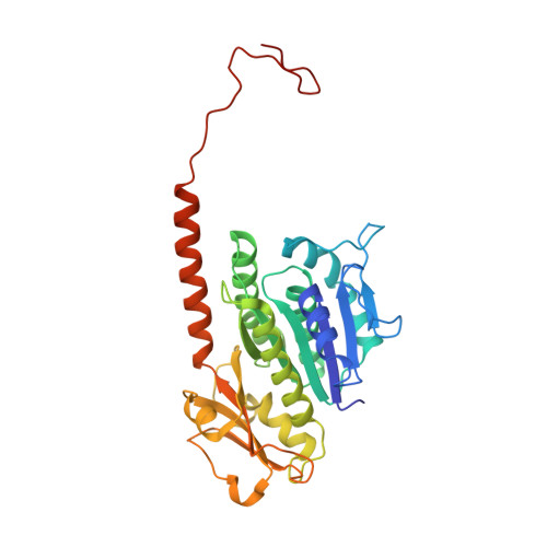
    3ZBP, 3ZBQ

  • PubMed Abstract: 

    Pseudomonas ΦKZ-like bacteriophages encode a group of related tubulin/FtsZ-like proteins believed to be essential for the correct centring of replicated bacteriophage virions within the bacterial host. In this study, we present crystal structures of the tubulin/FtsZ-like protein TubZ from Pseudomonas bacteriophage ΦKZ in both the monomeric and protofilament states, revealing that ΦKZ TubZ undergoes structural changes required to polymerise, forming a canonical tubulin/FtsZ-like protofilament. Combining our structures with previous work, we propose a polymerisation-depolymerisation cycle for the Pseudomonas bacteriophage subgroup of tubulin/FtsZ-like proteins. Electron cryo-microscopy of ΦKZ TubZ filaments polymerised in vitro implies a long-pitch helical arrangement for the constituent protofilaments. Intriguingly, this feature is shared by the other known subgroup of bacteriophage tubulin/FtsZ-like proteins from Clostridium species, which are thought to be involved in partitioning the genomes of bacteriophages adopting a pseudo-lysogenic life cycle.


  • Organizational Affiliation
    • MRC Laboratory of Molecular Biology, Francis Crick Avenue, Cambridge CB2 0QH, UK.

Macromolecule Content 

  • Total Structure Weight: 37.74 kDa 
  • Atom Count: 2,805 
  • Modeled Residue Count: 315 
  • Deposited Residue Count: 333 
  • Unique protein chains: 1

Macromolecules

Find similar proteins by:|  3D Structure
Entity ID: 1
MoleculeChains  Sequence LengthOrganismDetailsImage
PHIKZ039333Phikzvirus phiKZMutation(s): 0 
EC: 3.6.5
UniProt
Find proteins for Q8SDC3 (Pseudomonas phage phiKZ)
Explore Q8SDC3 
Go to UniProtKB:  Q8SDC3
Entity Groups
Sequence Clusters30% Identity50% Identity70% Identity90% Identity95% Identity100% Identity
UniProt GroupQ8SDC3
Sequence Annotations
Expand
Reference Sequence

Small Molecules

Ligands 1 Unique
IDChains Name / Formula / InChI Key2D Diagram3D Interactions
GDP

Query on GDP


Download:Ideal Coordinates CCD File
B [auth A]GUANOSINE-5'-DIPHOSPHATE
C10 H15 N5 O11 P2
QGWNDRXFNXRZMB-UUOKFMHZSA-N

Experimental Data & Validation

Experimental Data

  • Method: X-RAY DIFFRACTION
  • Resolution: 1.70 Å
  • R-Value Free:  0.203 (Depositor), 0.169 (DCC) 
  • R-Value Work:  0.168 (Depositor), 0.166 (DCC) 
  • R-Value Observed: 0.170 (Depositor) 
Space Group: P 1 21 1
Unit Cell:
Length ( Å )Angle ( ˚ )
a = 43.494α = 90
b = 66.243β = 112.69
c = 52.581γ = 90
Software Package:
Software NamePurpose
PHENIXrefinement
XDSdata reduction
SCALAdata scaling
PHASERphasing

Structure Validation

View Full Validation Report


Ligand Structure Quality Assessment 


Entry History 

Deposition Data

Revision History  (Full details and data files)

  • Version 1.0: 2013-04-03
    Type: Initial release
  • Version 1.1: 2013-04-10
    Changes: Database references
  • Version 1.2: 2013-06-12
    Changes: Database references
  • Version 2.0: 2018-01-24
    Changes: Atomic model, Source and taxonomy
  • Version 2.1: 2023-12-20
    Changes: Data collection, Database references, Derived calculations, Other, Refinement description